Download
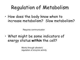
1 / 28
280 likes | 439 Views
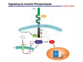
Regulation of inositol 1,4,5- trisphosphate (IP 3 ) receptors and IP 3 -induced Ca 2+ release. H. De Smedt K.U.Leuven, Belgium. IP 3 R1: 3D reconstruction (Hamada et al., 2003). N. N. IP 3 -binding domain. Bar: 10 nm. C. C. Regulatory domain. cytosol. ER. Transmembrane region.

E N D
Regulation of inositol 1,4,5- trisphosphate (IP3) receptors and IP3-induced Ca2+ release H. De Smedt K.U.Leuven, Belgium
IP3R1: 3D reconstruction (Hamada et al., 2003) N N IP3-binding domain Bar: 10 nm C C Regulatory domain cytosol ER Transmembrane region Ca2+ Luminal loop
Agonists IP3 Ca2+ Plasma membrane associated: Homer-mGluR TRP; RhoA-TRPC1 G; RACK-1 Cytoskeletal proteins: Actin; MyosinII Ankyrin; Tallin; Vinculin 4.1N Kinases and phosphatases: PKA; IRAG-PKG Fyn, BANK- PTK FKBP12-Calcineurin PP1 Cytosolic proteins: Calmodulin; CaBP IRBIT, CARP HAP1A-Htt Cyt C, Bcl-2, BclXL x x Caspase-3 ER Ca2+ Intraluminal proteins: Chromogranins; Calnexin N N C C
IP3R I II III + CaM [IP3] (µM) Part 1: Regulation of the IP3R by CaM IICR • CaM • (Michikawa et al., 1999) IP3R I II III + CaM (Missiaen et al., 1999) 300 [Ca2+] (nM)
Calmodulin-binding sites on IP3R1 Ca2+-indep. CaM R1:PPKKFRDCLFKLCPMNRYSAQKQFWKAAKPGAN R2:PPKKFRDCLFKVCPMNRYSAQKQYWKAKQAKQG R3:PPKKFRDCLFKVCPMNRYSAQKQYWKAKQTKQD W1577A (Zhang et al, 2001; Nosyreva et al, 2002) R1:LDSQVNNLFLKSHN-IVQKTAMNWRLSARN-AARRDSVLA R2:LDSQVNTLFMKNHSSTVQRAAMGWRLSARSGPRFKEALGG R3:LDAHMSALLSSGGSCSAAAQRSAANYKTATRTFPRVIPTA CaCaM 31 25 18 Cytosol 13 Endoplasmic reticulum
N 226 581 C IP3 binding core 100 80 [3H]IP3 binding (%) 60 40 20 Control Ca2+ CaM CaM1234 Ca2+/CaM1234 Ca2+/CaM 1 581 IP3 binding core suppressor Both Ca2+ and CaM bind to the suppressor and inhibit IP3 binding but CaM is not the Ca2+ sensor
600 CaM 400 Ca2+i (nM) 200 CaM1234 CaBP1 0 0 250 500 750 1000 IICR is inhibited by CaM, CaM1234 and CaBP1 Intact COS cells 0.5 µM 1 µM 100 µM ATP Control Time (s)
1-225 T1 1-225 T1 1-225 T1 1-225 T1 1-225 T1 1-225 T3 1-225 T3 1-225 T3 1-225 T3 1-225 T3 86 76 75 87 CaM-binding sites on the suppressor domain (199) (200) Bosanac et al., Mol. Cell, 2005
Exchange or deletion of the less conserved region in the arm subdomain of the suppressor is critical for IP3 sensitivity
1-225 T1 1-225 T1 1-225 T1 1-225 T1 1-225 T1 1-225 T3 1-225 T3 1-225 T3 1-225 T3 1-225 T3 CaM-binding sites on the suppressor domain CaM-binding CaM- and CaBP1-binding (199) (200) Bosanac et al., Mol. Cell, 2005
Depletion of CaM with high-affinity CaM-binding peptides uncouples IICR from IP3 binding
Ac-RRKEQKTGHAVRAIGRE-NH2 EGTA 0.5 µM Ca2+ Depletion of endogenous CaM by MLCK peptide inhibits IICR in permeabilized L15 fibroblasts 0.5 µM Ca2+ control Ac-RRKWQKTGHAVRAIGRL-NH2 (Kd 6 pM)
MLCK peptide does not change the affinity for IP3 Control Control 1 µM MLCK 10 µM CaM 10 µM MLCK
Inhibition of IICR by different CaM-binding peptidesis dependent on their affinity for CaM
IP3 MLCK pep CaM CaM CaM1234 The inhibition by MLCK peptide can be reversed by Ca2+-CaM but not by CaM1234
Regulation via CaM and Ca2+CaM sites IP3 Depletion of constitutive CaM CaCaM ? 31 25 Ca2+ sensor 18 Cytosol Ca2+ -indep CaM ATP NH2 P 1852S P P P ATP COOH ATP P 13 Endoplasmic reticulum
Part 2: The IP3R as a Ca2+-leak pathway;Truncation by caspase-3 Ca2+ -indep CaM Caspase-3 cleavage IP3 KKDDEVDRDA CaCaM 31 25 Ca2+ sensor 18 Cytosol ATP NH2 P P P P ATP COOH ATP P 13 Endoplasmic reticulum
Caspase-3 mediated cleavage of IP3R1in a DT40 triple(IP3R)-KO background Caspase-3 activity Apoptosis
The role of a leaky IP3R in mouse oocytes mouse eggs upon injection of channel only domain mRNA Aged mouse eggs Perturbed Ca2+ oscillations
Altered intracellular Ca2+ homeostasis in Alzheimer’s disease Role of presenilins (PS) Overexpression of PS1/PS2 mutants → potentiation IP3-mediated Ca2+ release → increased IP3 sensitivity → suppression of Capacitive Ca2+ Entry (CCE) PS1/PS2 knockouts → decreased IP3-mediatedCa2+ release → potentation of CCE Hypothesis: altered ER Ca2+ content? (LaFerla et al., Nature, 2003)
Presenilin (PS1/PS2) knockout decreases the [Ca2+]ER as measured by ER-targeted aequorin CaCl2 CaCl2 WT WT PS1-Rescue PSDKO How does Presenilin alter the [Ca2+ ]ER?
PS DKO Rescue WT IP3R1 IP3R3 IP3R1 IP3R3 Expression of proteins involved in Ca2+ handling PS DKO Rescue WT IP3R1 IP3R3 IP3R1 IP3R3 SERCA CRT BiP
WT PS DKO Rescue Basal Ca2+ leak is increased in PSDKO cells Intact cells Permeabilized cells
RNAi knock-down of IP3R1restored IICR in PSDKO cells to control values PS DKO RNAi control PS DKO RNAi PS DKO WT IP3R1 IP3R3 IP3-induced Ca2+ release vs A23187 WT PS DKO PS DKO PS DKO RNAi RNAi control
WT PS DKO PS DKO; IP3R1-RNAi IP3R1knock-down by RNAi restored Ca2+ content and Ca2+ leak in PSDKO cells to control values Ca2+ content (fmol/cell) PS DKO IP3R1-RNAi PS DKO RNAiControl WT PS DKO PS DKO RNAi control
The Ca2+ leak is not IP3 dependent Hyperphosphorylated IP3R (Oakes et al., PNAS 2005 ) or enhanced Bcl-2 expression are not responsible for leak in PSDKO cells wt PSDKO IP3R1 P-Ser wt PSDKO Bcl-2 Full size IP3R1 PS DKO PS DKO + heparin 95kDa There is significant cleavage of the IP3R with formation of a channel-only domain Why is IP3R1 ‘leaky’ ?
Part 2: The IP3R as an IP3-independent Ca2+-leak channel • Cleavage of the IP3R in a position upstream of the channel domain creates an IP3-independent Ca2+channel • The IP3R behaves as an IP3-independent Ca2+-leak channel in presenilin-knockout cells
Zerihun ASSEFAGeert BULTYNCKSarah KOCKSNael NADIF KASRI Karolina SZLUFCIKVeerle VANDERHEYDENLeen VERBERTBenoit DEVOGELAERE IP3-team (Leuven, Belgium) Jan B. PARYS - Ludwig MISSIAEN Humbert DE SMEDT In collaboration with the groups of:A. Galione (University of Oxford) K. Török (St George’s University of London) R.A. FISSORE (Univ. Massachusetts)M.J. BERRIDGE – M.D. BOOTMAN – L. RODERICK (Babraham) B. DE STROOPER (Genetics – K.U.Leuven) G. CALLEWAERT (Physiology – KULAK)