Download
1 / 23
250 likes | 861 Views
REGULATION OF RESPIRATION. outline. Theory Preparations before operation Operation in neck Item. Ⅰ Theory.

E N D
outline Theory Preparations before operation Operation in neck Item
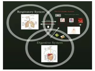
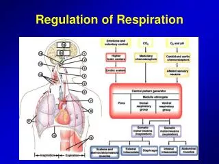
ⅠTheory The activity of the respiratory centers regulate the movement of respiration. The respiratory centre activity in turn is influenced by afferent impulses from the lungs and various other parts of the body. Factors which modify respiration are nervous factors and chemical factors (CO2 and O2 tensions or H+ in blood or cerebrospinal fluid). There are many methods of recording respiratory movement: directly recording the change in airway pressure and electricity in diaphragm. In this experiment we adopt the methods of recording diaphragm movement.
ⅠTheory To observe the effects of some factors on respiration and grasp the recording technical of respiration movement.
Ⅱ APPARATUS & AGENTS APPARATUS a. Mammal animal operating apparatus b. Tracheal cannula c. Syringe(30ml and 50ml respective) d. A rubble tube 50cm in length e. A bladder of ball filled with CO2 f. Protective electrode g. Power Lab System AGENTS: a. 20% urethane b. 0.9% saline solution c. 3% lactic acid
Ⅲ Preparations before operation • 1 Anesthesia: 20% urethane (5mg/kg) is administered through the marginal vein in the ear. The position where we begin to inject anesthetic should be near to distal of marginal ear as possible as we can. The velocity of injection should be slow and we should monitor the respiration of the animal.
Ⅲ Preparations before operation • 2 Fixing: After anesthesia, the rabbit is fixed. We should lay the rabbit on its back on the operation table, then fix the limbs with the help of the ropes.
Ⅲ Preparations before operation • 3 Shearing: Before the operation, the hair of the operation site should be sheared. This step we should avoid injuring the animal’s skin.
Ⅳ Operation in neck • 1 Slice the skin: With the scissors make a mid incision 3-4 centimeters in length through the skin over the region from the lower jaw to the breastbone. The anterior chest is now exposed.
Ⅳ Operation in neck • 2 Separate the subcutaneous tissue and muscle to expose the trachea In this step, we should avoid injuring the blood and nerve.
Ⅳ Operation in neck • 3 Endotracheal intubation Separate the trachea from other tissues, pass a thread under it, make a “⊥”shape incision on the trachea 2-3 cm under the throat, then insert the tracheal cannula toward the lung and tie the cannula and the trachea together.
Ⅳ Operation in neck • 4 Separate the vagus nerve Identify carefully the three nerves located in carotid sheath: vagus nerve is the thickest, depressor nerve is the thinnest and the sympathetic nerve is moderate. Isolate them by glass dissecting needle and pass various color threads separately under them for preparation.
Ⅳ Operation in neck The operating action of isolating blood vessels and nerves should be gentle by using glass dissecting needle. The isolating sequence is from thin to thick.
ⅤSeparate the xiphoid process • 1 Make a skin incision located in xiphisternum 2-3cm in length. • 2 Open the abdominal cavity along the linea alba abdominis 2-3cm in length. • 3 Separate the muscles in the middle of the belly to expose and isolate the xiphoid process.
ⅤSeparate the xiphoid process • 4 The peritoneum around the xiphoid process was sheared carefully. Lift the xiphoid process, you can see two slips of diaphragm adhering to the xiphoid process. • 5 Cut between the body of sternum and the xiphoid process.
ⅤSeparate the xiphoid process • 6 Connect the xiphoid process with the computer through a pressure transducer. Do not injury the diaphragm and avoid pneumothorax.
Ⅵ Item • (1) Observe the normal respiration movement. • (2) Increase the concentration of CO2 during inspiration:a ball filled with CO2 is joined with one side of trachea cannula, increasing the concentration of CO2 during inspiration via opening the clip of the bladder. When the obvious change of respiratory movement happened,close the clip instantly.
Ⅵ Item • (3) Observe the change of respiration movement when decrease the concentration of O2 during inspiration: close one side of the trachea cannula with a bladder filled with atmosphere through a bottle full of sodium-lime and have the animal breath in this bladder, then wait for a moment. The amount of carbon dioxide dose not change (because the sodium-lime absorbed the carbon dioxide during expiration)
Ⅵ Item • (4) Observe the change of respiration movement when enlarge the dead space: close one side of trachea cannula. Then wait for a moment, the other side of trachea cannula is connected with a rubber tube about 50 cm in length. The animal respire through the rubber tube. • (5) Observe the change of respiration movement when increase the H+ concentration in blood: about 1-2ml 3% lactic acid is intravenously injected rapidly.
Ⅵ Item • (6) Observe the change of respiration movement when pour gas in pulmonary: when the respiration is stable,the pulmonary is poured into 20 ml atmosphere via a 30ml syringe connected with one side of trachea at the end of inspiration. At the same time, the other side of treacha is blocked. • (7) Observe the change of respiration movement when cut vagus nerves. One side of vagus nerves or both side of that was cut.
Ⅶ Precautions • 1.We should keep the trachea clear to move blood blockage out. • 2.Make an mid line incision to isolate xiphisternum for fear that blood too much. • 3.Pay attention not to injury diaphragm to avoid pneumothorax. • 4.Insert the electrode into diaphragm appropriately . • 5.Dcreasing the CO2 concentration during inspiration should be slow. • 6.The dose of 3% lactic acid should not too much.
Ⅷ Question • 1.What is the difference of mechanism that enhancing respiratory movement by increasing CO2 concentration during inspiration or decrease the concentration of O2 during inspiration or increase the H+ concentration in blood? • 2.What is the role that vagus play in rhythmic respiration movement?