Vermeer—The Geographer
300 likes | 651 Views
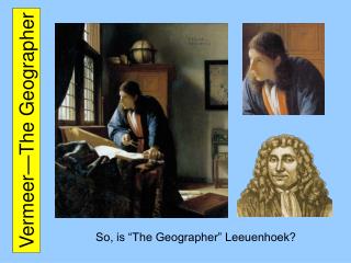
Vermeer—The Geographer. So, is “The Geographer” Leeuenhoek?. Relative Sizes. SEM. SEM. both. both. both. both. TEM. TEM. Simple Scope: Only One Lens (Advantage = Less Distortion). Leeuenhoek’s Microscope. View Through Light Microscope.

Vermeer—The Geographer
E N D
Presentation Transcript
Vermeer—The Geographer So, is “The Geographer” Leeuenhoek?
Relative Sizes SEM SEM both both both both TEM TEM
Simple Scope: Only One Lens (Advantage = Less Distortion) Leeuenhoek’s Microscope
View Through Light Microscope • [Top left] View (Elder pith) with Original Leeuwenhoek microscope. • [Top right] Modern Leitz optical microscope (light-ground). • [Bottom Left] Modern Leitz optical microscope (dark-ground). • [Bottom right] Cambridge Stereoscan electron microscope image.
Light Filter Light Through Compound Scope Compound Scope: More than One Lens (Advantage = Greater Magnification)
What is Resolution? • The object of microscopy is not just to increase magnification, but to do so while retaining sufficient resolution. • Resolution is the ability to see two items as two separate things, i.e., two dots as two separate dots. • The resolution a microscope is capable of achieving is the smallest distance between two dots such that the two dots may be observed (resolved) as separate entities. • In less technical terms, lower resolution means an increased degree of fuzziness, i.e., less focusable [sic?] specimens.
dark-field illumination oblique illumination bright-field illumination Bright- vs. Dark-Field (2/3) This image of the surface of a leaf shows the differences in contrast between these types of illumination. Bright-field illumination has very limited contrast. This image clearly shows that it is very useful to experiment with contrast techniques. Oblique illumination gives a relief-like enhancement of contrast. In 19th century microscopes there was often an arrangement for oblique illumination. Early microscopists knew that this technique has great advantages! Dark-field illumination is also one of the most rewarding techniques. Objects smaller than the resolving power of the objective can also made visible simply because they light up! It is thought that Antony van Leeuwenhoek observed bacteria using a kind of dark-field illumination! (see: http://www.microscopy-uk.org.uk/mag/artapr02/dark.html)
Bright- vs. Dark-Field (3/3) Section of Mouse Eye (above) Hydrodyction the water net
TEM of a freeze fracture replica of a mouse pancreas cell. Notice, the nuclear pores on the surface of the nucleus. Freeze Fracturing
Gram Staining Bacillus anthracis Escherichia coli
Acid-Fast Staining Mycobacterium avium complex (MAC) with acid fast stain often has the characteristic appearance shown here with numerous mycobacteria filling macrophages. Such macrophages may be distributed diffusely or in clusters.
Numerical Aperture Need photo of objective lens showing NA, etc. Need equation showing dependence of resolution of NA and wavelength.