Cells into Tissues
Cells into Tissues. Differentiating of cell types during development Germ Layers Endoderm Mesoderm Ectoderm. Endoderm. Endoderm. epithelial lining of the whole of the digestive tube lining cells of all the glands which open into the digestive tube

Cells into Tissues
E N D
Presentation Transcript
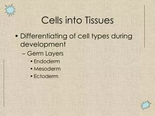
Cells into Tissues • Differentiating of cell types during development • Germ Layers • Endoderm • Mesoderm • Ectoderm
Endoderm • epithelial lining of the whole of the digestive tube • lining cells of all the glands which open into the digestive tube • the epithelium of the auditory tube and tympanic cavity; trachea, bronchi, and air cells of the lungs; urinary bladder and part of the urethra; and follicle lining of the thyroid gland and thymus. • stomach, colon, liver, pancreas, urinary bladder, lining of the urethra, epithelial parts of trachea, lungs, pharynx, thyroid, parathyroid, intestines.
Mesoderm • The formation of a mesoderm led to the development of a coelom. • Organs formed inside move, grow, and develop independently of the body wall while fluid cushions and protects them from shocks. • Forms skeletal muscle, skeleton, dermis of skin, connective tissue, urogenital system, heart, blood and vessels, spleen.
Ectoderm • Forms CNS, the lens of the eye, cranial and sensory, ganglia and nerves, pigment cells, head connective tissues, epidermis, hair, and mammary glands.
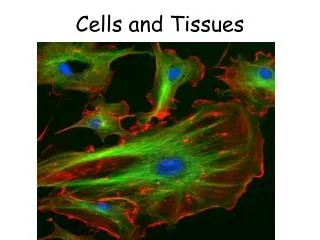
4 Types of Tissue • Epithelium • Connective • Muscle • Nervous
Tissues: groups of cells closely associated that have a similar structure and perform a related function • Four types of tissue • Epithelial = covering • Connective = support • Muscle = movement • Nervous = control • Most organs contain all 4 types • Connective tissue has non-living extra-cellular material (matrix) between its cells
Epithelial and Connective Tissues • Epithelial tissues • Classes • Junctions • Glands • Connective Tissues • Matrix • Cells • Types
EPITHELIAL TISSUES • Sheets of cells • Specialized contacts/cell junctions (see below) • Basal lamina: protein scaffolding secreted by epithelial cells • Basement membrane: reticular fibers (crossed collagen network) that supports epithelium--really associated connective tissue
EPITHELIAL TISSUES CON’T… • Connective tissue support • Nutrients from capillaries in underlying connective tissue • Nerves pass through • Easily regenerates • E.g. skin, lining of gut, mucous membranes
Simple: just one layer or cell shape Stratified: multiple layers and cell shapes Classes of Epithelia
Squamous E.g. epidermis Transitional epithelium E.g. urinary structures--bladder Stretches from 6 cells to 3 cells thick as bladder fills and expands Stratified Epithelia
Quiz!! E Can You Identify the Classes of Epithelium? D A B C
Cell Junctions • Desmosome: binding spots between cells with proteins called cadherins • Tight junctions: impermeable • E.g. gut tube, doesn’t let enzymes from gut into blood stream • Gap junctions: tubes that let small molecules pass between cells
Features of Apical Surface of Epithelium • Microvilli:(ex) in small intestine • Finger-like extensions of the plasma membrane of apical epithelial cell • Increase surface area for absorption • Cilia: (ex) respiratory tubes • Whip-like, motile extensions • Moves mucus, etc. over epithelial surface 1-way • Flagella:(ex) spermatoza • Extra long cilia • Moves cell
Features of Lateral Surface of Epithelium • Cells are connected to neighboring cells via: • Proteins-link cells together, interdigitate • Contour of cells-wavy contour fits together • Cell Junctions • Desmosomes-adhesive spots on lateral sides • Tight Junctions-at apical area, plasma membrane of adjacent cells fuse, nothing passes • Gap junction-spot-like junction occurring anywhere, lets small molecules pass
Features of the Basal Surface of Epithelium • Basal lamina: supportive sheet between epithelium and underlying connective tissue • Selective filter • Basement membrane = basal lamina plus underlying reticular fiber layer • Attaches epithelium to connective tissue below • Sometimes the two are used interchangeably
Name that Epithelial Feature!(name and location on cell) 3 • Cilia • Tight junction • Microvilli • Basement membrane 3 1 1 2 2 4 4
Glands: epithelial cells that make and secrete a water-based substance • Exocrine Glands • Secrete substance onto body surface or into body cavity • Have ducts • E.G., salivary, mammary, pancreas, liver • Endocrine Glands • Secrete product into blood stream • Either stored in secretory cells or in follicle surrounded by secretory cells • Hormones travel to target organ to increase response • No ducts
CONNECTIVE TISSUES • “Areolar tissue” as model • Connects skin to underlying structures • Universal in body • Underlies epithelium, supports capillaries, small. • Always originates from mesenchyme • CELLS in MATRIX
Extracellular matrix • Fibers • Collagen gives structure • Reticular fibers (crossed collagen) gives order • Elastin gives elasticity • Ground substance • Jelly-like material made of sugar-protein molecules (proteoglycans)
Cells of Connective Tissues • Fibroblasts make fibers • Immune cells in areolar tissue • Macrophages • Plasma cells • Mast cells • Neutrophils, Lymphocytes
“Loose” connective tissues • Adipose tissue mostly under skin and in mesenteries • Reticular: organized 3-D network of fibers that support lots of cells • E.g. marrow, spleen, lymph nodes
“Dense” Connective tissues • Irregular • Thick fibers running in many planes • E.g. dermis, fibrous capsules around organs • Regular • Aligned parallel fibers • Resists tension • E.g. tendon, ligaments, aponeuroses • Sometimes with elastic fibers (e.g. ligamentum nuchae)
Other Connective Tissues • Bone • Cartilage • Blood