Immune System
Immune System. Is a self / non-self recognition and achieved by having every cell display a marker based on the major histocompatibility complex. RBCs. Source of immune cells (Lymphoid organs). Primary organs: bone marrow and the thymus gland .

Immune System
E N D
Presentation Transcript
Immune System Is a self / non-self recognition and achieved by having every cell display a marker based on the major histocompatibility complex.
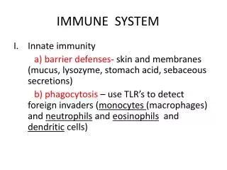
Source of immune cells (Lymphoid organs) • Primary organs: bone marrow and the thymus gland . • Secondary organs: Adenoids, tonsils, spleen , lymph nodes , Peyer's patches and the appendix. Surface barriers or mucosal immunity: 1. Skin. 2. Mechanically, pathogens are expelled from the lungs by coughing , sneezing and ciliary movement of respiratory cells. 3. Sticky mucus in respiratory and gastrointestinal tracts. 4. Saliva, tears and nasal secretions contain lysoenzyme antinfective agents. 5. Vaginal secretions are also slightly acidic . Spermine and zinc in semen destroy pathogens. Lactoperoxidase is a powerful antinfective enzyme found in mother's milk. 6. Highly acidity of stomach.
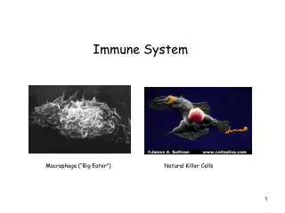
Kinds of phagocytes according to their origin: • Promonocytes made in bone marrow and released into the blood and called circulating monocytes. • Macrophages of liver called Kupffer cells. • Macrophages of brain called microglia. • Macrophages of kidney called mesoangial cells. Types Of Phagocytes : • Natural killer cells (large granular lymphocytes ) move in the blood and lymph and attach to the glycoproteins on the surfaces of infected virus and bacteria and kill them. • 2. Polymorphonuclear neutrophils, phagocytes that have no mitochondria and get their energy from stored glycogen. • 3. Eosinophils are attracted to cells coated with complement
Types of Immunity • Innate immunity: The innate immunity system is what we are born with and it is nonspecific; all antigens are attacked pretty much equally. Each immu ne cells conjugate with the antigen by pattern-rcognition receptors. • Acquired immunity: Occurred as a response to the infection and is of two types: A. Humoral immunity. B. Cell mediated immunity.
Cell mediated Immunity:T- cells originated from bone marrow lymphocyte which migrate to thymus gland to multiply and carry genetic information and tested for recognition and binding to antigen. Processing of T-cells occurs largely during foetal life and early childhood .There are three types of T-cells :A. Helper T-Cells: Secrete lymphokines that stimulate cytotoxic T cells and B cells to grow and divide , attract neutrophils and enhance the ability of macrophages to engulf and destroy microbes.B. Killer T-cells : Known as cytotoxic T-cells: Secrete lymphotoxins which cause cell lysis.C. Memory T-cells : Are programmed to recognize and respond to a pathogen once it has invaded.D. suppressor T cell : Suppresses the immune response of B cells and other T cells once the end of destroy the invaders.
2. Humoral Immunity:The humoral immune response involves a complex series of events after antigens enter the body. First, macrophages take up some of the antigen and attach it to class II MHC molecules, then bind the antigen to T helper cells which become stimulated and divide and secrete stimulatory molecules called interleukins. The interleukins activate any B lymphocytes to bind to the antigen. The activated B cells then divide and secrete antibodies. Finally, the secreted antibodies bind the antigen and help to destroy it.Antibodies:Antibodies are Y-shaped proteins called immunoglobulins (Ig) and are made only by B cellsCategorize antibodies into five main classes: IgM, IgG, IgA, IgD, and IgE.
The antibodies inactivate antigens by: (a)Complement fixation. (b)Neutralization. (c)Agglutination. (d)Precipitation.
Stem cells Some call them magic seeds, for their ability to replicate indefinitely and morphologically into any kind of tissue. Stem cells have traditionally been characterised as either embryonic (pluripotent) or tissue-specific (multipotent). Stem cells are the source of all cells - brain, skin, heart and others - that make up the human body. Just like a plant stem that branches into leaves and flowers, stem cells branch out to form different bits of our bodies.
Source of stem cells • Pre-implantation embryos. • Embryo from IVF. • The fluid that surrounds a developing baby in the womb pre-implantation embryos. • Embryo from IVF. • Fluid that surrounds a developing baby in the womb. • Umbilical cord. • Bone marrow cells (Haematopoietic & stromal stem cells).
Advantage of adult stem cells : Adult stem cells offer the opportunity to utilize small samples of adult tissues, to obtain an initial culture of a patient's own cells for expansion and subsequent implantation in the same person (Autologous transplant). This process avoids immune rejection by the recipient and also protects the patients from viral, bacterial or other contamination from another individual (donor) as in case of allogenic transplant. Disadvantages : • Culturing adult stem cells in-vitro is very difficult and has not been possible for some types. • They have a very short life, when cultured in-vitro as compared to embryonic cells.
Stem cells & Heart disease: Congestive heart failure results from loss or dysfunction of heart muscle cells. The disease afflicts 4.8 million people , with 400,000 new cases each year. The disease result from coronary heart disease , heart attack , the sudden close of the blood vessels supplying oxygen to the heart. Two major cells of the heart : • Cardiomyocyte that contracts to eject the blood out of the heart's main pumping chamber ( ventricle). • Vascular endothelial cells which form the inner lining of the blood vessel. • Smooth muscle cells which form the wall of the blood vessel. The heart has a large demand for blood flow, and these specialized cells are important for developing a new network of arteries to bring nutrients and oxygen to the cardiomyocytes after a heart has been damaged.
Stem cell therapy of heart failure : Injection of selected bone marrow cells with a high capacity to develop into cells of multiple types ( haematopoietic stem cells). When these cells injected into the damaged wall of the ventricle, these cells led to the formation of the of new cardiomyocytes , vascular endothelium, and smooth muscle cells. Thus generating de novo myocardium , including coronary arteries, arterioles, and capillaries. The newly formed myocardium occupied 68 percent of the damaged portion of the ventricle nine days after the bone marrow cells were transplanted. The partial repair of the damaged heart muscle suggest that the transplanted haematopoietic stem cells respond to signals in the environment near the injured myocardium. • Vasculogenesis: Is the in situ differentiation of mesodermal precursors to angioblasts that differentiate into endothelial cells to form the primitive capillary network. Vasculogenesis is limited to early embryogenesis and is believed not to occur in the adult. • Angiogenesis : the sprouting of new capillaries from the preexisting blood vessels and occurs in both the developing embryos and postnatal life.
Neuronal stem cells Every sensation, action and thought explains the complicated processes of the central nervous system (CNS), which consists of the brain and the spinal cord. The brain is the central computer of our body interpreting outside information and controlling every action. The spinal cord connects the brain with the rest of the body by sending out millions of electrical signals. Neuronal cells are responsible for receiving and processing every piece of information the brain sends the rest of the body. Neurons are made up of four parts—the cell body which houses the nucleus and most of the cell organelles, dendrites, an axon, and axon terminals. Dendrites are bush like projections that bring information from other neurons to the cell body. The axon, a longer projection, sends information away form the cell body. Injuries of spinal cord is irreversible and cause paralysis and the information from brain and other regions of the body are blocked. This disease affects many millions of people around the world .
Defects of spinal cord injury led to : • Swelling causes additional damage to the spinal cord as pressure builds in the confined space between the cord and vertebrae as a result of scar tissue that builds up around the area of injury which blocks the neurons from reconnecting once the cord has been severed. • Swelling cuts off the blood supply to the neurons and glial cells which intern lead to additional neuronal cell death and migration of more immune cells to the injury site.
Stem cell therapy: Reconnection must be reestablished and activate new neurons and glial cells to regenerate and replace the injured ones. Once nerve cells were damaged they were gone, eliminating hope for complete recovery from paralysis. Scientists recently discovered that new neurons in specific regions of the adult mammalian brain . Neural stem cells were isolated from the dentate gyrus of the hippocampus and the walls of the ventricular system called the ependymal layer. The progeny of these stem cells differentiate in the granule cell layer, meaning neurogenesis continues late into adult rodent life. These stem cells also migrate along the rostral migratory stream to the olfactory bulb, where they differentiate into neurons and glial cells .
Derived undifferentiated embryonic stem cells (ES cells) from fetal spinal cord tissue and then mature them into cells that are suitable to implant into the damaged spinal cord. When using ES cells, researchers have two options: they can treat ES cells, allowing them to mature into CNS cells in vitro before transplantation, or they can directly implant differentiated cells and depend on signals from the brain mature the cells. • Treating injured spinal cord of rats with undifferentiated embryonic stem cells (ES cells) from fetal spinal cord tissue led to marked differentiation of it , filling the area normally occupying by glial scarring. After five weeks the stem cells had migrated further away from the implantation site. Although a number of them had died, there was still enough for the rats to have a growing supply of neurons and glial cells. Most of the surviving cells were oligodendrocytes and astrocytes, but some neurons were found in the middle of the cord. The rats regained limited use of their legs. Paralysis had been cured!!