Download
1 / 15
180 likes | 750 Views
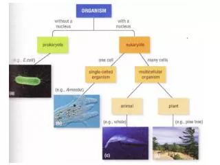
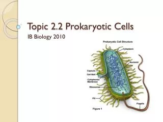
2.2 Prokaryotic Cells. 2 .2 Prokaryotic Cells. Draw and label a diagram of the ultrastructure of Escherichia coli (E. Coli) as an example of a prokaryote Use images of bacteria as seen in electron micrographs to show the structure.

E N D
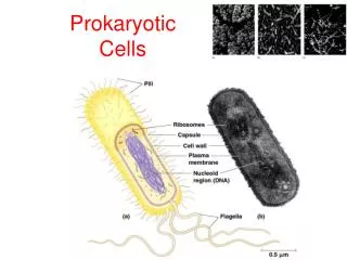
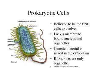
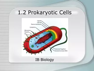
2.2 Prokaryotic Cells Draw and label a diagram of the ultrastructure of Escherichia coli (E. Coli) as an example of a prokaryote • Use images of bacteria as seen in electron micrographs to show the structure. • The diagram should show the cell wall, plasma membrane, mesosome, cytoplasm, ribosomes, plasmid, pili, flagella and nucleoid.
2.2 Prokaryotic Cells Annotate your diagram with the functions each named structure: • cell wall – forms a protective outer layer that prevents damage from outside and bursting if internal pressure is too high • plasma membrane – controls entry and exit of substances, pumping some of them in by active transport
mesosome – increases the area of membrane for ATP production. May move the DNA to the poles during cell division • cytoplasm – contains enzymes that catalyse the chemical reactions of metabolism and DNA in a region call the nucleoid. Site for metabolic reactions • flagella - structures protruding from the cell wall with a corkscrew shape, using energy, they can be rotated, to propel the cell from on area to another unlike eukaryotic flagella, they are solid and inflexible, working like a propeller
ribosomes7Os– synthesize proteins by translating messenger RNA. (Some proteins stay in the cell and others are secreted • nucleoid– contains naked DNA which stores the genetic information that controls the cell and is passed on to daughter cells • pili - protein filaments protruding from the cell wall used for cell to cell adhesion when bacteria stick together to form aggregations of cells when two cells are exchanging DNA during conjugation
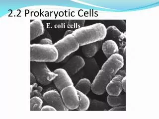
Plasmids -Extra-nucleoid DNA . Plasmids can self-replicate particularly before binary fission. They are associated with conjunction which is horizontal gene transfer. • Identify structures from an electron micrographs of E. coli shown on the next slide.
Reproduction of prokaryotic cells by binary fission • Prokaryotic cells divide by binary fission. • This is an asexual method of reproduction in which a 'parental' cell divides into two smaller but equally sized cells. The cells are genetically identical and form the basis of a reproductive clone.
Review Questions: Q. 1 How do prokaryotic cells divide? A. By mitosis B. By meiosis C. By budding D. By binary fission Q.2 Which function is carried out by the flagellum of the prokaryote? A. Movement of food towards the cell B. Movement of the whole cell from one place to another C. Movement of naked nucleic acid inside the cell D. Movement of water around the cell to speed up gas exchange
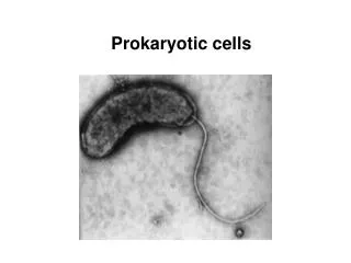
Questions 3 and 4 refer to the following micrograph of an E.coli bacterium undergoing reproduction.
Q. 3 The scale bar represents 0.5 µm. How long are both cells in total? A. 5.0 x 10- 6 m B. 5.0 x 10- 9 m C. 2.5 x 10- 6 m D. 2.5 x 10- 9 m Q. 4 In the diagram what does label X identify? A. Nucleoid region B. Chromatin C. Histones D. Endoplasmic reticulum
Q. 5 (b) Distinguish between prokaryotic cells and eukaryotic cells. [6] (b) Draw and label a diagram of the ultrastructure of Escherichia coli. [6] (c) Annotate your diagram in (5) (b) above with functions of each part labelled. [6]