Download
1 / 28
280 likes | 292 Views
Learn about the cellular structure, mitosis, and anatomical terminology. Understand the different microscope techniques used in biological research.

E N D
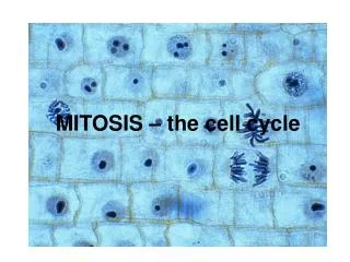
Bio211 - Laboratory 1 Microscope Cellular Structure Cell Cycle Mitosis
Anatomical Terminology Know these terms Anatomical Position– body standing erect, facing forward, upper limbs at the sides, palms facing forward Material starting here will be tested on LECTURE Exam 1
Body Sections Know this
Anatomical Subdivisions Used most in clinical situations Used most in surgical/anatomical study situations
Body Regions Know the terms on this slide and their locations on the body END of material for LECTURE EXAM 1 Fig 2.3 in Lab Manual
“Seeing” in Biology • There are many different tools that biologists/anatomists can use to ‘see’ biological samples at high resolution. Some include: • Light microscope (2-D)* • Electron microscope* • Transmission electron microscopy (2-D) • Scanning electron microscopy (3-D) • Confocal laser scanning microscope (optical sections through a 3-D specimen; good for 3-D) • Atomic force microscope (one of the most powerful tools for determining the surface topography of native biomolecules at subnanometer resolution)
Resolving Power Need a refresher on the metric system? See our course Web site in the “Study Guides and Helpers” Section From: http://www.mih.unibas.ch/Booklet/Lecture/Chapter1/Chapter1.html
Light Microscope (Erythrocytes) Nucleus 7.5 m (Use as a guide to size)
Transmission Electron Micrograph RBC Two-dimensional representation of a 3-D object From: http://www.upei.ca/~morph/webct/Modules/EM/EM.html
The Scanning Electron Microscope No, although the hairdo is similar, this is NOT your instructor!! From: http://www.mih.unibas.ch/Booklet/Lecture/Chapter1/Chapter1.html
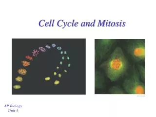
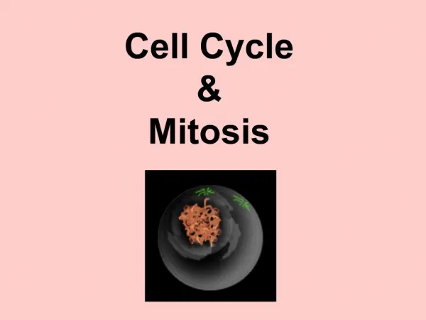
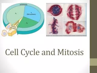
The Cell Cycle • series of changes a cell undergoes from the time it forms until the time it divides • stages • interphase • mitosis • cytoplasmic division • differentiation G0 Differentiatedcells may spend all their time in G0 (neurons, skeletal muscle, red blood cells) Stem cells may never enter G0
The Cell Cycle Must Have Controls • If DNA is damaged, cell must NOT be allowed to enter mitosis • DNA must be completely replicated before mitosis takes place • At metaphase, the chromosomes must be correctly positioned at the spindle fiber equator • Each phase of the cell cycle must be completed before the next is begun • DNA/Cell replication must not proceed unless a ‘signal to proceed’ is received
What are the Controls of the Cell Cycle? • cell division capacities vary greatly among cell types • skin and bone marrow cells divide often • liver cells divide a specific number of times then cease • chromosome tips (telomeres) that shorten with each mitosis provide a mitotic clock (cell senescence) • cells divide to provide a more favorable surface area to volume relationship • growth factors and hormones stimulate cell division • hormones stimulate mitosis of smooth muscle cells in uterus • epidermal growth factor stimulates growth of new skin • contact inhibition • Cyclins and Cyclin-dependent kinases provide central control • tumors are the consequence of a loss of cell cycle control
The Cell Cycle and Mitosis • Review from Biology… • What is the cell cycle? Why does mitosis happen? INNKEEPER, POUR ME ANOTHER TEQUILA!
The Cell Cycle and Mitosis • INNKEEPER (INTERPHASE) • POUR (PROPHASE) • ME (METAPHASE) • ANOTHER (ANAPHASE) • TEQUILA (TELOPHASE/CYTOKINESIS)
Prophase What structure joins the sister chromatids together?
Plant Mitosis (Allium root tip) Prophase Telophase Interphase Anaphase Metaphase Late anaphase /early telophase Images from: http://biology.about.com/od/mitosis/ig/Mitosis-Image-Gallery/index.htm
Reminders about using the microscope… • Reminders for using microscope • Start at low power and locate a promising area to view at higher power, center the object of interest, THEN go to higher power • When you focus on an area under higher power, be sure to move the FINE focus up and down slightly to be sure you see everything you need to see • Try and use both eyes (for a binocular scope) • Review and USE the instruction sheet for Lab 1 in your Lab Guide that was handed out today!!
Cellular Structure • This should be a review from General Biology • Use your textbook if necessary to label the composite cell Figure 5.5 (page 60) in your lab Manual. • List a function for each of the cellular components you labeled (see page 59 Lab Manual).
The Cell Be able to label a figure like this (figure 5.5, page 60 in Lab Manual) Give a function for each organelle
The Cell Cycle and Mitosis • Be sure you are able to recognize… • Each phase of mitosis • For each of the phases of mitosis you are required to look at both: • Whitefish blastula • Allium root tip • Use the photographs in Wood’s Lab Manual to guide you • If you are still in doubt about something you see, just ASK!
Lab Safety/Rules • Please be sure to REVIEW the laboratory rules in your Laboratory Guide and SIGN the sheet on the bench in the front of the lab BEFORE YOU LEAVE TODAY. • If you EVER have a question about something in the lab, always ASK before ACTING! • There is NO FOOD OR DRINK allowed in the Science Department laboratories • Open-toed shoes are not allowed in the Science labs
What you should do in lab today… • Refer to the Instructions for Laboratory 1 in your Laboratory Guide. If you think you have a question – READ IT AGAIN, THEN ASK! • Before you leave the lab today… • Call me over to your lab table so I can check if your group can recognize the different mitotic figures • Sign the Lab Safety Rules sign up sheet Remember to try the Online Quizzes (on gserianne.com Web site) -AFTER you think you have mastered the material for a lab - and/or before the Lab Exam
For next lab… • Epithelial and Connective Tissue • Read Exercises 7 and 8 in Wood’s Lab Manual • Look at the histological photos in your Lab Manual and in your textbook (Ch. 4, Sec 1 and 2) • Label the diagrams in your Lab Manual