Download
1 / 54
550 likes | 932 Views
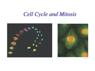
Cell Cycle and Mitosis. Honors Biology. 100 µm. (a) Reproduction. An amoeba, a single-celled eukaryote, is dividing into two cells. Each new cell will be an individual organism (LM). Figure 12.2 A. Why is Cell Division Important?. Unicellular organisms

E N D
Cell Cycle and Mitosis Honors Biology
100 µm (a) Reproduction. An amoeba, a single-celled eukaryote, is dividing into two cells. Each new cell will be an individual organism (LM). Figure 12.2 A Why is Cell Division Important? • Unicellular organisms • Reproduce by cell division increasing the population.
200 µm 20 µm (b) Growth and development. This micrograph shows a sand dollar embryo shortly after the fertilized egg divided, forming two cells (LM). (c) Tissue renewal. These dividing bone marrow cells (arrow) will give rise to new blood cells (LM). Why Do Multicellular Organisms Depend on Cell Division? • Development & Growth • Repair (ex: tissue renewal) • Maintenance
Cell Division (aka Mitosis) • Makes 2 genetically identicaldaughter cells from 1 parent cell • Before cells divide • They duplicate their genetic material ensures that each daughter cell receives an exact copy of the genetic material, DNA
What is the structure of a chromosome? • Where in a cell is the genetic material/chromosomes located? • Nucleus • Chromatin is an uncoiled mass of DNA and histone proteins • Exists in this form the majority of the time! • Histones are proteins that help DNA condense • As a cell prepares to divide it coils up/condenses: • We call this CHROMOSOMES (condensed DNA)
DNA Molecules • DNA (in nucleus of eukaryotes) can be in 2 forms • Chromatin: DNA is not tightly packed together (loosely coiled) • Occurs during interphase • Chromosomes : tightly packed together (TIGHTLY coiled) • Occurs during mitosis (cell division)
Genes • Segments of DNA (that make up the chromosome) are called genes • A gene is a piece/segment of DNA that stores genetic information
What happens to chromosomes during cell division? • What needs to be done to a chromosome before it can divide? • It must DUPLICATE! (DNA Replication) • After duplication each chromosome consists of 2 identically joined copies Sister Chromatids • Sister Chromatids are held together by centromeres Chromatid Sister Chromatids (condensed, duplicated chromosome) Chromatin
Double Chromosome Structure Kinetochoreattaches to spindle fibers Sister
Chromosomes • Every eukaryotic species has a characteristic, unique # of chromosomes in EACH cell nucleus • Ex: Humans have 46 chromosomes • # of chromosomes does NOT necessarily equal complexity
The Cell Cycle • The mitotic phase alternates with interphase in the cell cycle • Interphasemitosisinterphasemitosis
What is Mitosis!? • Mitosis is the process where cells divide to produce new cells - Occurs in healing (Ex: if you cut yourself) • New cells are also produced as you grow - Ex: Day-to-day life (new skin cells!) • ALL eukaryotic organisms produce new cells through mitosis
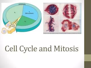
Cell Cycle • Consists of 2 broad stages • 1. Growing Stage called Interphase • 2. Cell Division called Mitotic Phase (M Phase) • The majority of the cell cycle (90%) is spent in Interphase
INTERPHASE S(DNA synthesis) G1 CytokinesisMitosis G2 MITOTIC(M) PHASE Figure 12.5 Phases of the Cell Cycle
Interphase can be divided into subphases • G1 phase (GAP 1 phase) • cell grows in size • varies most in length from cell to cell • S phase (synthesis phase) • DNA is copied (DNA replication) • Single Double • Each chromosome is single • DNA replication occurs • Chromosomes have doubled each consisting of two sister chromatids • G2 phase (GAP 2 phase) • More growth and preparation (make proteins) for mitosis
Mitotic Phase • After Interphase, Mitotic Phase begins • Two parts of M Phase: • Mitosis (division of the nucleus) 2) Cytokinesis (division of the cytoplasm)
M Phase • Mitosis – the nucleus and duplicated chromosomes divide and create two identical daughter cells • Cytokinesis – the process by which the cytoplasm is divided in two. • Cytokinesis usually begins before Mitosis is completed.
Refresher……. The Cell Cycle: G1 phase: Growth S phase: DNA replication G2 phase: Preparation for cell division M phase: Mitosis and Cytokinesis
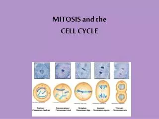
Remember…. Interphase Prophase Metaphase Anaphase Telophase Cytokinesis IPMATC IPassedMy Accelerated Tough Class
Interphase chromosome • Consists of G1, S, G2 • Occurs BEFORE Mitosis begins • During S phase, the cell copies its DNA • Chromosomes appear as threadlike coils • Made of Chromatin, a combination of DNA and protein molecules • As the cell prepares to divide, its chromatin fibers condense, becoming the compact structure we call a chromosome. • Chromosomes are copied (# doubles) Condensed, duplicated chromosome
Interphase • Each chromosome has now been condensed and duplicated and consists of 2 sister chromatids • The region where the two chromatids are joined tightly together is called the centromere.
Mitosis • Continuous pathway (Early, Mid, & Late) • Consists of 4 phases and cytokinesis • Prophase • Metaphase • Anaphase • Telophase • Cytokinesis
Prophase (X’s)“Pack Together” First phase of Mitosis: • Chromatin becomes tightly coiled = chromosomes • DNA “packs” together • Spindle Fibers (made by thecentrioles) begins to form in the cytoplasm 3. Nuclear envelope breaks down Late:Nucleus and nucleolus disappear
Prophase: Centrioles Spindles
Prophase: 2. Centrioles move DNA supercoils into chromosomes
Metaphase (X’s)“Meet in the Middle” Second phase of Mitosis: • Chromosomes attach to the spindle at the centromeres • Chromosomes line up in the middle of cell • Called equatorial or metaphase plate • Spinder fibers pull and tug chromosomes to line up
Anaphase (V’s)“Adios and Away” Third phase of Mitosis: • Spindle pulls apart chromosomes • SISTER CHROMATIDS separate athe the centromere and begin moving to opposite ends (poles) of the cell • Each chromatid is now considered its own chromosome
***Remember that each chromatid has the same DNA so each is now its own chromosome*** Anaphase:
Telophase (V’s)“Two New Cells” Fourth phase of Mitosis: • Chromosomes reach end of spindle • Spindle breaks down (disappear) • Cleavage furrow begins to form 4. Nuclear membrane begins to reform 5. 2 daughter nuclei 6. Chromosomes chromatin
Telophase: Spindle fall apart Cleavage furrow
Cytokinesis“Division of the Cytoplasm” • Occurs in Late telophase • In animal cells • a cleavage furrow forms, which pinches the cell in two. • In plant cells • produce a cell plate at the middle of the cell • At the end of cytokinesis, there are two distinct IDENTICAL daughter cells.
Cytokinesis • Final Phase of Cell Division/M Phase • Cleavage furrow pinches all the way through • Result is two new cells • 2 cells then enter Mitosis begins again! • G1, S, G2 (Interphase) • PMAT & Cytokinesis • Each new cell at the end of mitosis is DIPLOID • has a full set of chromosomes
Cleavage furrow 100 µm Contractile ring of microfilaments Daughter cells (a) Cleavage of an animal cell (SEM) Figure 12.9 A Cytokinesis: A Closer Look • In animal cells • Cytokinesis occurs by a process known as cleavage, forming a cleavage furrow
Vesiclesforming cell plate Wall of patent cell 1 µm Cell plate New cell wall Daughter cells Figure 12.9 B (b) Cell plate formation in a plant cell (SEM) • In plant cells, during cytokinesis • A cell plate forms
PROMETAPHASE G2 OF INTERPHASE PROPHASE Aster Centrosomes(with centriole pairs) Fragmentsof nuclearenvelope Kinetochore Early mitoticspindle Chromatin(duplicated) Centromere Nonkinetochoremicrotubules Kinetochore microtubule Nucleolus Chromosome, consistingof two sister chromatids Figure 12.6 Nuclearenvelope Plasmamembrane
METAPHASE ANAPHASE TELOPHASE AND CYTOKINESIS Metaphaseplate Cleavagefurrow Nucleolusforming Nuclear envelopeforming Daughter chromosomes Centrosome at one spindle pole Spindle Figure 12.6
2 3 5 1 4 Chromatinecondensing Nucleus Chromosome Nucleolus Metaphase. The spindle is complete,and the chromosomes,attached to microtubulesat their kinetochores, are all at the metaphase plate. Prophase. The chromatinis condensing. The nucleolus is beginning to disappear.Although not yet visible in the micrograph, the mitotic spindle is staring to from. Prometaphase.We now see discretechromosomes; each consists of two identical sister chromatids. Laterin prometaphase, the nuclear envelop will fragment. Telophase. Daughternuclei are forming. Meanwhile, cytokinesishas started: The cellplate, which will divided the cytoplasm in two, is growing toward the perimeterof the parent cell. Anaphase. Thechromatids of each chromosome have separated, and the daughter chromosomesare moving to the ends of cell as their kinetochoremicrotubles shorten. Figure 12.10 Mitosis in a plant cell
Cell Cycle and Mitosis Animations • http://www.ucopenaccess.org/courses/APBiologyI/course%20files/multimedia/lesson17/lessonp.html • http://highered.mcgraw-hill.com/olcweb/cgi/pluginpop.cgi?it=swf::535::535::/sites/dl/free/0072437316/120073/bio14.swf::Mitosis%20and%20Cytokinesis • http://www.sumanasinc.com/webcontent/animations/content/mitosis.html • http://www.johnkyrk.com/mitosis.html