Download

1 / 54
540 likes | 705 Views
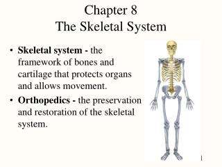
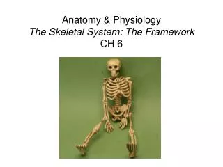
Bones support and give shape to the body. This framework helps protect vital organs and furnishes attachment points for muscles, ligaments and tendons. Bones also store minerals and contain hematopoietic bone marrow.

E N D
Bones support and give shape to the body. This framework helps protect vital organs and furnishes attachment points for muscles, ligaments and tendons. Bones also store minerals and contain hematopoietic bone marrow.
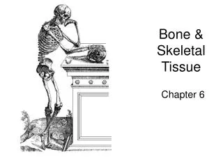
Bone is a specialized form of connective tissue containing about 50% solid matter and 50% water.
Bone consists of a hard outer shell called compact bone and an inner spongy structure called cancellated or cancellous bone
The bone surfaces are covered by a tough fibrous vascular membrane called the periosteum.
The long bones grow in length at the ‘epiphysis’ (the ends of the developing bones)
Diaphysis (shaft) Medullary cavity epiphysis
Bones are classified according to their shape.LongFlatShort Irregular
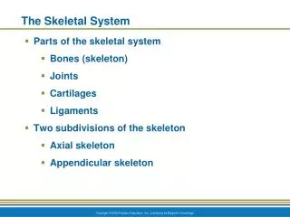
The skeleton is divided into two main parts:the axial: including the skull, vertebral column, ribs and sternumthe appendicular: including the limbs
The skull includes two major segments the cranium (brain case) and the facial bones.All skull bones are immobile except the mandible.The skull bones are united by sutures. Within the bones of the skull are hollows called sinuses. The function of the sinuses include, lessening the bone weight, providing chambers for vacalization and moistening and warming air.
Facial bone Facial bone
Vertebrae Vertebral AnatomyThe horse has 37 vertebrae:* Cervical (neck)- 7 * Thoracic (chest) - 18* Lumbar (lower back) - 6 (except in Arabs - 5) * Sacral (pelvis) - 5 (fused) The dog has 31 vertebrae:* Cervical (neck)- 7 * Thoracic (chest) - 13 * Lumbar (lower back) - 7 * Sacral (pelvis) - 3 (fused) The cat has 31 vertebrae:* Cervical (neck)- 7 * Thoracic (chest) - 13 * Lumbar (lower back) - 7 * Sacral (pelvis) - 3 (fused)
C-1 and C-2 are called the atlas and the axis. The words atlas (holding up the world) and axis (what the world spins on) come from Greek mythology. There can be an instability in this area in large dogs that will cause neurologic problems. The cervical vertebrae are quite flexible, for obvious reasons.
As the cervical vertebrae become the thoracic vertebrae they go past the shoulder (S). The nerves that come off this cervical-thoracic junction at the shoulder are called the brachial plexus (you cannot see nerves on a plain radiograph). They innervate the front legs on each side. Each of the thoracic vertebrae corresponds to a rib (R) on each side of the chest
As we continue down the thoracic vertebrae you can visualize how high their dorsal spinal processes are. Also notice how these processes start to get smaller as we get closer to the lumbar vertebrae
Moving towards the end of the thoracic vertebrae we come to what is termed the thoracolumbar (T-L) junction. It is a very common area to have VSC disease. As we pass into the lumbar vertebrae we have now made our way into the lower back
The 7 lumbar vertebrae eventually lead into the sacral vertebrae (S). The fused sacral vertebrae are hard to visualize because they are within the pelvis. After the sacrum we are at the tail
Clavicle (collarbone)feline: a small flat bone attached to the scapulacanine: a vestigial bone that is not attached to any other bone and may be absent in some dogs.
Humerusthe long bone extending from the shoulder to the elbowthe humerus articulates with the scapula and the radius/ulna
Radius/UlnaUlna: the caudle bone that articulates with the humerus at the olecranonRadius: the cranial bone that articulates with the humerus and ulna at the elbow
Carpuscomposed of 7 – 8 irregularly shaped bones in two rows. This joint is called the wrist in humans
Pelvis illeum ichium pubis
Femur femur
Tibia/Fibula tibia fibula
A joint is an articulation between bones, or between bones and cartilage. They are classified by the degree of movement they permit
Synarthrosis: allow no movement Fibrous tissue
Amphiarthrosis allow slight movement Cartilaginous tissue
Diarthroses freely permit movement Synovial joints Hinge Ball and socket Gliding Pivot Condyloid
Synovial membrane: lines the joint capsule and secretes synovial fluid