human cardiomyocytes
30 likes | 44 Views
Cardiomyocytes are derived from the whole heart (of a single donor) that has been dissociated into single cells and cultured using differential adhesion. Learn more from https://www.creative-bioarray.com/Human-Cardiomyocytes-CSC-C2847-item-39324.htm

human cardiomyocytes
E N D
Presentation Transcript
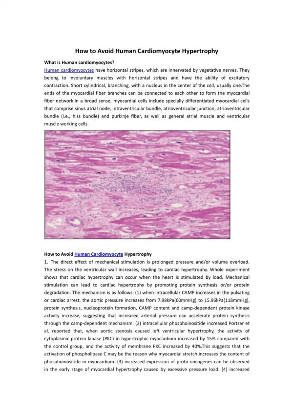
How to Avoid Human Cardiomyocyte Hypertrophy What is Human cardiomyocytes? Human cardiomyocytes have horizontal stripes, which are innervated by vegetative nerves. They belong to involuntary muscles with horizontal stripes and have the ability of excitatory contraction. Short cylindrical, branching, with a nucleus in the center of the cell, usually one.The ends of the myocardial fiber branches can be connected to each other to form the myocardial fiber network.In a broad sense, myocardial cells include specially differentiated myocardial cells that comprise sinus atrial node, intraventricular bundle, atrioventricular junction, atrioventricular bundle (i.e., hiss bundle) and purkinje fiber, as well as general atrial muscle and ventricular muscle working cells. How to Avoid Human Cardiomyocyte Hypertrophy 1. The direct effect of mechanical stimulation is prolonged pressure and/or volume overload. The stress on the ventricular wall increases, leading to cardiac hypertrophy. Whole experiment shows that cardiac hypertrophy can occur when the heart is stimulated by load. Mechanical stimulation can lead to cardiac hypertrophy by promoting protein synthesis or/or protein degradation. The mechanism is as follows: (1) when intracellular CAMP increases in the pulsating or cardiac arrest, the aortic pressure increases from 7.98kPa(60mmHg) to 15.96kPa(118mmHg), protein synthesis, nucleoprotein formation, CAMP content and camp-dependent protein kinase activity increase, suggesting that increased arterial pressure can accelerate protein synthesis through the camp-dependent mechanism. (2) intracellular phosphoinositide increased Portzer et al. reported that, when aortic stenosis caused left ventricular hypertrophy, the activity of cytoplasmic protein kinase (PKC) in hypertrophic myocardium increased by 15% compared with the control group, and the activity of membrane PKC increased by 40%.This suggests that the activation of phospholipase C may be the reason why myocardial stretch increases the content of phosphoinositide in myocardium. (3) increased expression of proto-oncogenes can be observed in the early stage of myocardial hypertrophy caused by excessive pressure load. (4) increased
expression of actin and myosin genes when cultured myocardial cells were continuously stimulated by stretch, the expression of -mhc and -actin genes increased.(5) other calcium ion channels, sodium ion influx and intracellular alkalinity ratio can play important regulatory roles in myocardial hypertrophy induced by stretch stimulation. 2. Chemical stimulation of humoral factors may also promote cell hypertrophy or proliferation. (1), norepinephrine (NE), animal experiments show that the long-term injection of hypertension dose NE may induce myocardial hypertrophy, the myocardial hypertrophy may mainly through alpha 1 - R works, some scholars also will join the rest of the myocardial cell culture can be found that myc gene transcription is 5 ~ 10 times, and promote myocardial hypertrophy, the reaction can be non-specific, alpha 1 receptor antagonist blocking can strengthen by protein kinase C, activator PNA increase NE by alpha receptors, activation of phosphatidyl inositol, protein kinase C system, activation of oncogene expression. (2) androgen Cabral, etc in rats to induce neurogenic hypertension after baroreceptor nerve found that male rats significantly higher than that of females, left ventricular weight/body weight ratio to testosterone can cause similar male neurogenic hypertension left ventricular myocardial hypertrophy in rats, and give estradiol can inhibit left ventricular weight increase, its mechanism is still not clear, may be related to cancer gene. (3) angiotensin Ⅱ (Ang Ⅱ) Ang Ⅱ AT1 and AT2 receptors points two subtypes, AT1 receptor and myocardial positive inotropic and chronotropic action and the growth of the myocardial cell hypertrophy related to Ang Ⅱ add to the myocardial cell culture, c fos, c - jun, c - mye protocarcinogenic gene expression, such as strengthening rapidly, and lead to increased protein synthesis, the induced myocardial hypertrophy.Ang Ⅱ can also cause angiotensin original gene and transforming growth factor beta 1, prompting the myocyte hypertrophy, the reaction can be Ang Ⅱ AT1 receptor blockers block, can also be PKC, prompt Ang Ⅱ by AT1 receptor activation inositol phosphate ester - protein kinase C system, activates protocarcinogenic gene expression. (4) endothelin (ET) was isolated from pig aortic endothelial cells by Yangisawa ET al in 1988.ET plays a role in regulating cell proliferation by binding to ET receptors on target cells.ET receptors are divided into ETA, ETB, ETC. Cardiovascular muscle cells are rich in ETA, and ETA can cause myocardial hypertrophy, possibly by increasing the expression of light bond 2, alpha - actin, troponin, and heavy chain mRNA of alpha and beta myosin.It has been reported that ETA may also play a role in myocardial hypertrophy caused by NE in the body.(5) growth hormone (GH) and insulin-like growth factor (TGF)Antonio et al. observed the effects of GH and TGF1 on the cardiovascular system of rats, and found that the myocardium was the target organ of GH and igf-1, and exogenous GH and igf-1 could cause hypertrophy of the myocardium of normal adult rats.The increased volume and pressure load can enhance the gene expression of cardiac igf-1.GH and igf-1 may also function indirectly through insulin metabolism or the adrenalin system.(6) interleukin 6(il-6) and mast cell factor (ct-1)keiko et al. found that when myocardial cells were stimulated by hypoxia, reperfusion and other factors, they could secrete a large amount of il-6, which was related to myocardial hypertrophy.Il-6, as a ligand, binds to the il-6 receptor to form homodimer GP130, which activates the tyrosine kinase and a series of reactions such as ras-paf-map kinase, promoting the transcription activity of cell genes.Ct-1 is a protein with molecular weight of 21.5KDa isolated from the supernatant of mouse embryonic stem cells during differentiation induction.It has been reported that cardiomyocytes also produce ct-1 and also affect intracellular signaling through GP130.The role of CT - 1 stimulate myocyte hypertrophy than Ang Ⅱ and ET were stronger, CT - 1 May also make the myocardial cell c fos, c - jun and ANP,
mRNA expression increased, that CT - 1 gene transcription activation and nerve cells. About Creative Bioarray Creative Bioarray is an innovative biotechnology company whose mission focuses on developing unique technologies that provide global scientists with high quality products and satisfactory services to facilitate the investigation of life science researches. We provide a wide range of high quality normal human and animal cells, cell culture medium and reagents, FISH probes, tissue arrays, microorganisms and equipments. In addition, we also offer series of related services including cell services, biosample services and histology services for the researcher to make their project better and faster.