Download
1 / 76
1.08k likes | 2.44k Views
Chapter 28: The Reproductive Systems. Male RPS. Male reproductive system. Gonads – testes Produces sperm and secretes hormones System of ducts – transport and stores sperm, assists in their maturation, and conveys them to the exterior

E N D
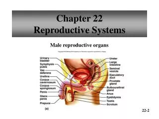
Male reproductive system • Gonads – testes • Produces sperm and secretes hormones • System of ducts – transport and stores sperm, assists in their maturation, and conveys them to the exterior • Epididymis, ductus deferens, ejaculatory ducts, and urethra • Accessory sex glands – adds secretions to semen • Seminal vesicles, prostate, and bulbourethral glands • Supporting structures • Scrotum supports testes and penis delivers sperm into female reproductive tract
Scrotum • Supporting structure for testes • Raphe – external median ridge • Scrotal septum – internally divides scrotum into two sacs, each with a single testis • Made up of subcutaneous layer and dartos muscle • Associated with each testis is the cremaster muscle • Normal sperm production requires a temperature 2-3°C below core body temperature • Cremaster and dartos muscle contracts or relaxes
Testes or testicles • Paired oval glands in the scrotum • Develops near kidney and descends through inguinal canals near 7th month of fetal development • Tunica vaginalis partially covers testes • Tunica albuginea – internal to tunica vaginalis • Extends inward forming septa that divide testis into lobules • Each of 200-300 lobules contains 1-3 seminiferous tubules • Sperm produced here through spermatogenesis
Seminferous tubule cells • Spermatogenic cells – sperm-forming cells • Spermatagonia (stem cell) develop from primordial germ cells that arise in yolk sac and enter testes in 5th week of development • Primary spermatocytes →secondary spermatocytes → spermatids → sperm cells → lumen • Sertoli cells or sustenacular cells– support cells • Tight junction form blood-testis barrier – prevents immune response against sperm cell surface antigens • Nourish spermatocytes, spermatids and sperm, phagocytize excess spermatid cytoplasm, control movements of spermatogenic cells, release sperm into lumen, produce fluid for sperm transport, secrete inhibin, regulate effects of testosterone and follicle-stimulating hormone (FSH) • Leydig (interstitial) cells found in spaces between seminiferous tubules • Secrete testosterone
Spermatogenesis • Takes 65-75 days • Begins with spermatogonia – diploid (2n) • Stem cells undergo mitosis to replace themselves and some continue development • Primary spermatocytes – diploid (2n) • Each duplicates its DNA and meiosis begins • Meiosis I – homologous pairs line up, crossing over occurs • Secondary spermatocytes (haploid or n) • 2 cells at end of Meiosis I • Each chromosome made up of 2 chromatids attached at centromere • Meiosis II – 2 chromatids separate • Spermatids – 4 haploid cells at end of meiosis II • Cells remain attached to each other by cytoplasmic bridges • Spermiogenesis – development of spermatids into sperm • Spherical spermatids transform into elongated sperm • Acrosome and flagella form, mitochondria multiply • Sertoli cells dispose of excess cytoplasm • Spermiation – release from connections to Sertoli cells • Not yet able to swim
PROPHASE METAPHASE ANAPHASE TELOPHASE PROPHASE II METAPHASE II ANAPHASE II TELOPHASE II no interphase between nuclear divisions (2n) (n) Crossing over occurs between homologues. Homologous pairs align randomly. Homologues separate from their partner. Chromosomes align at spindle equator. Sister chromatids of chromo-somes seperate. (n)
Possible ChromosomeCombinations 1 2 3 or or or
Sperm • Each day about 300 million sperm complete spermatogenesis • Head • Nucleus with 23 chromosomes (haploid or n) • Acrosome – vesicle filled with oocyte penetrating enzymes • Tail • Neck – contains centrioles forming microtubules that comprise remainder of tail • Middle piece – contains mitochondria • Principal piece – longest portion of tail • End piece – terminal, tapering portion of tail • Once ejaculated, sperm do not survive more than 48 hours in female reproductive tract http://newswatch.nationalgeographic.com/2013/03/19/sperm-works-best-in-the-winter/?utm_source=Facebook&utm_medium=Social&utm_content=link_fb20130326ngnw-sperm&utm_campaign=Content
Hormonal control of testes • At puberty, secretion of gonadotropin-releasing hormone (GnRH) increases • Stimulates anterior pituitary to increase secretion of luteinizing hormone (LH) and follicle-stimulating hormone (FSH) • LH stimulates Leydig cells to secrete testosterone • Synthesized from cholesterol mainly in testes • Suppresses secretion of LH and GnRH via negative feedback • Enzyme 5 alpha-reductase converts testosterone into dihydrotestosterone (DHT) in external genitals and prostate • FSH acts indirectly on spermatogenesis • FSH and testosterone act on Sertoli cells to stimulate secretion of androgen-binding protein (ABP) • ABP binds testosterone keeping concentration high • Testosterone stimulates spermatogenesis • Sertoli cells release inhibin which inhibits FSH
Androgens (testosterone and DHT) • Prenatal development • Testosterone stimulates male pattern of development or reproductive system ducts and descent of testes • DHT stimulates development of external genitalia • Development of male sexual characteristics • At puberty, they bring about development of male sex organs and development of male secondary sexual characteristics • Development of sexual function • Androgens contribute to male sexual behavior, spermatogenesis and sex drive (libido) • Stimulation of anabolism • Stimulate protein synthesis – heavier muscle and bone mass in men
Reproductive system ducts in males • Ducts of testis • Pressure generated by fluid produced by Sertoli cells push sperm along seminiferous tubules into straight tubules, rete testis, efferent ducts in epididymis and then ductus epididymis • Epididymis • Consists of tightly coiled ductus epididymis • Stereocilia are microvilli that reabsorb degenerated sperm • Site of sperm maturation – acquire motility and ability to fertilize • Can store sperm for several months • Continues as ductus (vas) deferens • Ductus (vas) deferens • Conveys sperm during sexual arousal through peristaltic contractions • Can also store sperm several months
Male reproductive system ducts • Spermatic cord • Ascends out of scrotum • Consists of ductus deferens as it ascends through scrotum, testicular artery, veins that drain testes and carry testosterone, autonomic nerves, lymphatic vessels, and cremaster muscle • Spermatic cord and ilioinguinal nerve pass through inguinal canal • Ejaculatory ducts • Formed by union of duct from seminal vesicle and ampulla of ductus deferens • Terminate in prostatic urethra • Eject sperm and seminal vesicle secretions just before release of semen into urethra • Urethra • Shared terminal duct of reproductive and urinary systems • Subdivided into prostatic urethra, membranous urethra, and spongy (penile) urethra • Ends at external urethral orifice
Accessory sex glands – secrete most of liquid portion of semen • Seminal vesicles - About 60% of semen volume • Secrete alkaline, viscous fluid containing fructose, prostaglandins, and clotting proteins (different from blood) • Prostate - About 25% of semen volume • Secretes milky, slightly acidic fluid containing citric acid, several proteolytic enzymes, seminalplasmin (antibiotic) • Bulbourethral glands • Secrete alkaline fluid that protects passing sperm by neutralizing acids from urine in urethra of the male • Mucus lubricates end of penis and lining of urethra
Semen and Penis • Semen • Mixture of sperm and seminal fluid • Typical volume 2.5-5 mL with 50-150 million sperm/mL • Slightly alkaline pH of 7.2-7.7 due to seminal vesicle secretions • Provides transport medium, nutrients, and protection • Coagulates after ejaculation due to clotting proteins??? • Penis • Contains urethra • Passageway for ejaculation of semen and excretion of urine • Body of penis – 3 cylindrical masses of tissue with erectile tissue • Glans penis – terminal opening is external urethral orifice • Prepuce or foreskin covers glans in uncircumcised men • Root of penis is attached portion • Erection – parasympathetic fibers release and cause local production of nitric oxide (NO) causing smooth muscle in arterioles to relax and dilate allowing large amounts of blood to enter penis
Female reproductive system • Gonads – ovaries • Uterine (fallopian) tubes or oviducts • Uterus • Vagina • External organs – vulva or pudendum • Mammary glands
Ovaries • Paired glands homologous to the testes • Produce • Gametes – secondary oocytes that develop into mature ova (eggs) after fertilization • Hormones including progesterone, estrogens, inhibin and relaxin • Series of ligaments hold ovaries in place • Broad ligament – part of parietal peritoneum • Ovarian ligament – anchors ovaries to uterus • Suspensory ligament – attaches ovaries to pelvic wall
Relative positions of the ovaries, the uterus, and supporting ligaments
Histology of ovary • Germinal epithelium – covers surface of ovary • Does not give rise to ova – cells that arise form yolk sac and migrate • Tunica albuginea • Ovarian cortex • Ovarian follicles and stromal cells (fibroblast-like cells) • Ovarian medulla • Contains blood vessels, lymphatic vessels, and nerves • Ovarian follicles – in cortex and consist of oocytes in various stages of development • Surrounding cells nourish developing oocyte and secrete estrogens as follicle grows • Mature (graafian) follicle – large, fluid-filled follicle ready to expel secondary oocyte during ovulation • Corpus luteum – remnants of mature follicle after ovulation • Produces progesterone, estrogens, relaxin and inhibin until it degenerates into corpus albicans
Oogenesis and follicular development • Formation of gametes in ovary • Oogenesis begins before females are born • Essentially same steps of meiosis as spermatogenesis • During early fetal development, primordial (primitive) germ cells migrate from yolk sac to ovaries • Germ cells then differentiate into oogonia – diploid (2n) stem cells • Before birth, most germ cells degenerate – atresia • A few develop into primary oocytes that enter meiosis I during fetal development • Each covered by single layer of flat follicular cells – primordial follicle • About 200,000 to 2,000,000 at birth, 40,000 remain at puberty, and around 400 will mature during a lifetime
Follicular development • Each month from puberty to menopause, FSH and LH stimulate the development of several primordial follicles • Usually, only one reaches ovulation/month • Primordial follicles develop into primary follicles • Primary oocyte surrounded by granulosa cells • Forms zona pellucida between granulosa cells and primary oocyte • Stromal cells begin to form theca folliculi • Primary follicles develop into secondary follicles • Theca differentiates into theca interna secreting estrogens and theca externa • Granulosa cells secrete follicular fluid in antrum • Innermost layer of granulosa cells attaches to zona pellucida forming corona radiata
Follicular development • Secondary follicle becomes mature (graffian) follicle • Just before ovulation, diploid primary oocyte completes meiosis I • Produces 2 unequal sized haploid (n) cells – first polar body is discarded and secondary oocyte • At ovulation, secondary oocyte expelled with first polar body and corona radiata • If fertilization does not occur, cells degenerate • If a sperm penetrates secondary oocyte, meiosis II resumes • Secondary oocyte splits into 2 cells of unequal size – second polar body (also discarded) and ovum or mature egg • Nuclei of sperm cell and ovum unite to form diploid zygote
Uterine (fallopian) tubes or oviducts • Provide a route for sperm to reach an ovum • Transport secondary oocytes and fertilized ova from ovaries to uterus • Infundibulum ends in finger-like fimbriae • Produce currents to sweep secondary oocyte in • Ampulla – widest longest portion • Isthmus – joins uterus • 3 layers • Mucosa – ciliary conveyor belt, peg cells provide nutrition to ovum • Muscularis – peristaltic contractions • Serosa – outer layer
Relationship of the uterine tubes to the ovaries, uterus, and associated structures
Uterus • Anatomy • Fundus, body, isthmus, and cervix (opens into vagina) • Normal position is anteflexion – anterior and superior over bladder (Cervix at right angle to vagina) • Ligaments maintain position – broad, uterosacral, cardinal and round • Histology – 3 layers • Perimetrium – outer layer • Part of visceral peritoneum – laterally the broad ligament. • Myometrium • 3 layers of smooth muscle • Contractions in response to oxytocin from posterior pituitary
Uterus Cont. • Endometrium – inner layer • Highly vascularized • Stratum functionalis –lines cavity, sloughs off during menstruation • Stratum basalis – permanent, gives rise to new stratum functionalis after each menstruation • Blood supply • Uterine arteries, arcuate arteries, radial arteries • Just before branches enter endometrium divide into • Straight arterioles supplying stratum basilis • Spiral arterioles supplying stratum functionalis change markedly during menstrual cycle • Cervical mucus - produced by secretory cells of cervix mucosa • Water, glycoproteins, lipids, enzymes, and inorganic salts • More hospitable to sperm near ovulation – thinner, more alkaline • Supplements energy needs of sperm, protect sperm from phagocytes and hostile environment of tract