Download
1 / 38
410 likes | 540 Views
Explore the latest advancements in gallbladder carcinoma, focusing on risk factors, geographical variations, and molecular pathogenesis. Learn about key insights for early detection and treatment options.

E N D
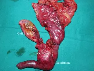
CARCINOMA GALL BLADDER-An Update Dr Sanjay De Bakshi MS; FRCS (Eng; Edin -ad eundem) Division of Surgical Gastroenterology Calcutta Medical Research Institute
PREAMBLE • Gallbladder cancer is the most common malignant tumour of the biliary tract worldwide . • It is also the most aggressive cancer of the biliary tract with the shortest median survival from the time of diagnosis. • This poor prognosis is due, in part, to an aggressive biologic behaviour and a lack of sensitive screening tests for early detection - resulting in delayed diagnosis and presentation at an advanced stage. C. H. E. Lai and W. Y. Lau, “Gallbladder cancer—a comprehensive review,” Surgeon, vol. 6, no. 2, pp. 101–110, 2008. X. Zhu, T. S. Hong, A. F. Hezel, and D. A. Kooby, “Current management of gallbladder carcinoma,” The Oncologist, vol. 15, no. 2, pp. 168–181, 2010. U. Dutta, “Gallbladder cancer: can newer insights improve the outcome?” Journal of Gastroenterology and Hepatology, vol. 27, no. 4, pp. 642–653, 2012.
INCIDENCE • Gallbladder carcinoma is the fifth most common gastrointestinal tumour. • Well- to moderately differentiated adenocarcinoma accounts for the most common form of gallbladder carcinoma. Gallbladder Carcinoma Update: Multimodality Imaging Evaluation, Staging, and Treatment Options. Alessandro Furlan et al American Journal of Roentgenology. 2008;191: 1440-1447
RISK FACTORS Adapted from -Gallbladder Cancer in the 21st Century. Rani Kanthan, Jenna-Lynn Senger, Shahid Ahmed, and Selliah Chandra Kanthan Journal of Oncology; Volume 2015 (2015),
RISK FACTORS • Demographic factors: (a)advanced age, (b)female gender, (c)obesity, (d)geography: South American, Indian, Pakistani, Japanese, and Korean, (e)ethnicity: Caucasians, Southwestern Native American, Mexican, and American Indians, (f)genetic predisposition.
RISK FACTORS • Gallbladder pathologies/abnormalities: (a) cholelithiasis, (Kaushik 2001; Rustagi and Dasanu 2014) (b) porcelain gallbladder, (Hundal and Schaffer 2014) (c) gallbladder polyps, (d) congenital biliary cysts *, (e) pancreaticobiliary maljunction anomalies. In Todani 's series of 154 cancers associated with bile duct cysts, 62 were in the gall-bladder, 1 in the liver and 2 in the pancreas.*
RISK FACTORS • Gallbladder pathologies/abnormalities: • The issue of microlithiasis • Gallbladder metaplastic changes appear to be more frequent in cases of micro-lithiasis and seem to be associated with a chronic thickening of the gallbladder wall. • Further studies are needed to evaluate a possible role of prophylactic cholecystectomy in this population to prevent the long term evolution of these early changes to cancerous lesions. Metaplastic Changes in Chronic Cholecystitis: Implications for Early Diagnosis and Surgical Intervention to Prevent the Gallbladder Metaplasia-Dysplasia-Carcinoma Sequence. Charalampos Seretis et al; J Clin Med Res. 2014 Feb; 6(1): 26–29.
RISK FACTORS • Chronic inflammation associated with (a)Primary sclerosing cholangitis (Bernstein 2001) (b)Ulcerative colitis (Bernstein 2001)
RISK FACTORS • Infections (a)Liver flukes (R. Hundal and E. A. Shaffer 2014), (b)Chronic Salmonella typhi and paratyphi infections (Nath 1997; Shukla 2000; Randi 2006; Nagaraja 2014) and (c)Helicobacter infection (Matsukura 2002; Kobayashi 2005).
RISK FACTORS • Exposures (a)Ingestion of certain medications (eg, oral contraceptives, INH, methyldopa) can increase the risk of gallbladder cancer. (b)Likewise, certain chemical exposures (eg, pesticides, rubber, vinyl chloride) and (c)Occupational exposures associated with working in the textile, petroleum, paper mill, and shoemaking industries increase the risk of gallbladder cancer. (d) Smoking (e)Exposures through water pollution (organopesticides, eg, dichlorodiphenyltrichloroethane and benzene hexachloride); heavy metals (eg, cadmium, chromium, lead); and radiation exposure (eg, radon in miners) are associated with gallbladder cancer.
PATHOGENESIS • Gallbladder cancer may arise in the gallbladder’s fundus (60%), body (30%), or neck (10%). • The development of gallbladder cancer is proposed to occur over a span of 5–15 years, with tissue alterations including metaplasia, dysplasia, carcinoma in situ, and invasive cancer K. S. Lim, C. C. Peters, A. Kow, and C. H. Tan, “The varying faces of gall bladder carcinoma: pictorial essay,” Acta Radiologica, vol. 53, no. 5, pp. 494–500, 2012. R. Hundal and E. A. Shaffer, “Gallbladder cancer: epidemiology and outcome,” Clinical Epidemiology, vol. 6, no. 1, pp. 99–109, 2014.
MOLECULAR PATHOGENESIS • Biologic pathways • Two distinct pathways proposed:- (1) a dysplasia-carcinoma sequence arising from metaplastic epithelium and (2) an adenoma-carcinoma sequence
MOLECULAR PATHOGENESIS • Genetic mutations • The genetic mutagenesis of Gall Bladder Carcinoma is ill understood. • Genes that have been studies • Oncogenes • Tumour Suppressor genes • Adhesion molecules and mucins • Angiogenesis • Cell cycle regulators • Apoptosis inducers
IMAGING • Sonography has a relatively high sensitivity for the detection of tumor at advanced stages, it is limited in the diagnosis of early lesions and is unreliable for staging. • Therefore, CT and, increasingly, MRI are more widely used for further characterization of potentially malignant gallbladder lesions and metastatic survey. Gallbladder Carcinoma Update: Multimodality Imaging Evaluation, Staging, and Treatment Options. Alessandro Furlan et al American Journal of Roentgenology. 2008;191: 1440-1447
IMAGING • On sonography, CT, or MRI, the presence of a large gallbladder mass that nearly fills or replaces the lumen, often directly invading the surrounding liver parenchyma, is highly suggestive of gallbladder carcinoma. Gallbladder Carcinoma Update: Multimodality Imaging Evaluation, Staging, and Treatment Options. Alessandro Furlan et al American Journal of Roentgenology. 2008;191: 1440-1447
IMAGING • On sonography, heterogeneous, predominantly hypoechoic tumor fills much or all of the gallbladder lumen. Gallbladder Carcinoma Update: Multimodality Imaging Evaluation, Staging, and Treatment Options. Alessandro Furlan et al American Journal of Roentgenology. 2008;191: 1440-1447
IMAGING • Primary gallbladder carcinoma is usually hypodense on unenhanced CT, with up to 40% of lesions showing hypervascular foci of enhancement equal to or greater than that of liver after IV contrast administration Gallbladder Carcinoma Update: Multimodality Imaging Evaluation, Staging, and Treatment Options. Alessandro Furlan et al American Journal of Roentgenology. 2008;191: 1440-1447
IMAGING • On MRI, gallbladder carcinoma usually shows hypo- to isointense signal characteristics on T1-weighted and moderately hyperintense signal characteristics on T2-weighted sequences Axial fast spin-echo T2-weighted MR image shows hyperintense mass (arrow) occupying gallbladder lumen and extending into adjacent liver parenchyma (arrowheads) with similar signal intensity. Gallbladder Carcinoma Update: Multimodality Imaging Evaluation, Staging, and Treatment Options. Alessandro Furlan et al American Journal of Roentgenology. 2008;191: 1440-1447
IMAGING • Gallbladder carcinoma may present as focal or diffuse asymmetric wall thickening in 20–30% of cases Gallbladder Carcinoma Update: Multimodality Imaging Evaluation, Staging, and Treatment Options. Alessandro Furlan et al American Journal of Roentgenology. 2008;191: 1440-1447
IMAGING • The initial detection of gallbladder carcinoma as a polypoid lesion occurs in 15–25% of cases. • Malignant lesions are usually larger than 1 cm in diameter and may have a thickened implantation base . Gallbladder Carcinoma Update: Multimodality Imaging Evaluation, Staging, and Treatment Options. Alessandro Furlan et al American Journal of Roentgenology. 2008;191: 1440-1447
IMAGING • Xanthogranulomatouscholecystitits presents usually with a • Symmetric thickening of the wall, • Continuous contrast—enhanced mucosal line. • Hypoattenuating nodules These represent abscesses or foci of inflammation. • Absence of any infiltration of surrounding tissues http://www.radiologyassistant.nl/en/p43a0746accc5d/gallbladder-wall-thickening.html
IMAGING Xanthogranulomatous Cholecystitis Masquerading as Gallbladder Cancer: Can It Be Diagnosed Preoperatively? Ashwin Rammohan, Sathya D. Cherukuri, Jeswanth Sathyanesan, Ravichandran Palaniappan, and Manoharan Govindan; Gastroenterology Research and Practice Volume 2014 (2014), Article ID 253645,
IMAGING • PET-CT in the assesment of Gall Bladder cancers. • Of value in detecting unsuspected distant metastases. Impact of integrated positron emission tomography and computed tomography on staging and management of gall bladder cancer and cholangiocarcinoma. Petrowsky et al J Heptol 2006 Jul; 45: 43-50. Clinical usefulness of 18-EDG PET-CT for patients with gall bladder cancer and cholangiocarcinoma. J Gastroenterol. 2010 May;45(5): 560-6.
IMAGING • Contrast-enhanced ultrasonography with perflubutane has been described in which gallbladder cancer shows continuous staining throughout the tumour and an “eruption sign” The efficacy of contrast-enhanced harmonic endoscopic ultrasonography in diagnosing gallbladder cancer. Mitsuru Sugimoto et al Sci Rep. 2016; 6: 25848.
IMAGINGContrast Enhanced US Scan Upper Left- Polypoid Upper Middle- Thick wall type Upper Right- Mass forming type Lower Left- Scattered blood vessels Lower Middle- Linear blood vessels Lower Right- Linear blood vessels Contrast-Enhanced Ultrasound in the Diagnosis of Gallbladder Diseases: A Multi-Center Experience. Lin-Na Liu, Hui-Xiong Xu et al October 31, http://dx.doi.org/10.1371/journal.pone.0048371
ROLE OF ADJUVANT THERAPY • Currently (2014), no adjuvant therapy that has been agreed upon as standard of care. Williams et al; Defining the role of adjuvant therapy; cholangiocarcinoma and gall bladder cancer. Semin Radial Oncol 2014 Apr; 24(2):94-104
ROLE OF NEO-ADJUVANT THERAPY • Of the 38 pts 33patients were treated with Gem-P (oxaliplatin, cisplatin; cetuximab-Gem-P) based therapy and 5 pts received chemoradiotherapy (CTRT) with wkly gemcitabine 300mg/m2. • Site of disease was • liver in 22 pts, • nodal in 12, • adjacent organ in 9 & • other in 3 pts.. • Neoadjuvant therapy in Indian patients with locally advanced gall bladder cancer: Tata Memorial Centre (TMC) experience. BhawnaSirohi, VipulSheth, Ashish Singh, Reena Engineer, MuktaRamadvar, Mahesh Goel, Shailesh V. Shrikhande2014 Gastrointestinal Cancers Symposium
ROLE OF NEO-ADJUVANT THERAPY • Response rate to NA therapy was • 5 (13%) complete response (CR), • 17(45%) partial response (PR), • 9 stable disease (SD), • 5 (13%) progressive disease and • not assessed in 2 patients (1 pt died post CTRT and 1 was inoperable at surgery). • Neoadjuvant therapy in Indian patients with locally advanced gall bladder cancer: Tata Memorial Centre (TMC) experience. BhawnaSirohi, VipulSheth, Ashish Singh, Reena Engineer, MuktaRamadvar, Mahesh Goel, Shailesh V. Shrikhande2014 Gastrointestinal Cancers Symposium
ROLE OF NEO-ADJUVANT THERAPY • Overall clinical benefit rate (CR+PR+SD) was 82%. • Of the 24 pts who underwent surgery, • 21 (87%) had curative resection and • 3 were inoperable. • Of 9 pts with SD, 6 received 2nd –line NA therapy as they were not downstaged enough to undergo surgery– 4 CTRT, 1 gemcitabine-cape, 1 cape-Ox. • Perioperative morbidity (biliary leak) was higher post CTRT. Overall, 7 pts have relapsed. • Neoadjuvant therapy in Indian patients with locally advanced gall bladder cancer: Tata Memorial Centre (TMC) experience. BhawnaSirohi, VipulSheth, Ashish Singh, Reena Engineer, MuktaRamadvar, Mahesh Goel, Shailesh V. Shrikhande2014 Gastrointestinal Cancers Symposium
ROLE OF NEO-ADJUVANT THERAPY • This is the first report of the use of neoadjuvant chemotherapy in patients with LA GB cancer. • CONCLUSION- preoperative chemotherapy is feasible with acceptable toxicity and perioperative morbidity. • Neoadjuvant therapy in Indian patients with locally advanced gall bladder cancer: Tata Memorial Centre (TMC) experience. BhawnaSirohi, VipulSheth, Ashish Singh, Reena Engineer, MuktaRamadvar, Mahesh Goel, Shailesh V. Shrikhande2014 Gastrointestinal Cancers Symposium
ROLE OF ADJUVANT & NEO-ADJUVANT THERAPY • Kaplan–Meier analysis demonstrates that neither neoadjuvant (a log rank p=0.59) nor adjuvant (b log rank p=0.16) therapy is associated with an improved probability of survival. • However, adjuvant chemotherapy is associated with a significant decrease in survival (b p=0.04) M. D. Anderson Cancer Center.
ROLE OF ADJUVANT & NEO-ADJUVANT THERAPY • Kaplan–Meier analysis demonstrates that neither neoadjuvant (a log rank p=0.59) nor adjuvant (b log rank p=0.16) therapy is associated with an improved probability of survival. • However, adjuvant chemotherapy is associated with a significant decrease in survival (b p=0.04) M. D. Anderson Cancer Center.
ROLE OF ADJUVANT & NEO-ADJUVANT THERAPY • Early surgical resection of biliary tract malignancies with 1 cm tumor-free margins or segmentectomy with lymph node dissection provides the best probability for long-term survival. • Currently available neo-adjuvant or adjuvant therapy does not improve survival. • Neither neoadjuvant nor adjuvant therapy increases survival after biliary tract cancer resection with wide negative margins. Glazer ES et al. J GastrointestSurg. 2012 Sep;16(9):1666-71. M. D. Anderson Cancer Center.
TARGETED THERAPIES IN GALL BLADDER CANCER • Common mutations reported in gallbladder cancer are KRAS (10%–67%), EGFR (63%), BRAF (0% to 33%), and erbB2/HER2 (16%–64%). • Early data suggest possible benefit from blockade of the epidermal growth factor receptor by the oral tyrosine kinase inhibitor erlotinib or anti-EGFR monoclonal antibody cetuximab. • Vascular endothelial growth factor (VEGF) is overexpressed in biliary tract cancers and has been proposed as a therapeutic target. The efficacy of bevacizumab, a monoclonal antibody targeting VEGF, in combination with erlotinib was assessed in a phase II trial showed onlty a modest benefit.
PROGNOSIS • A disease of very poor prognosis. • Surgery, the only curative therapy. (<20% operable) • Techniques of early diagnosis evolving. • Regional nodal status and the depth of tumor invasion (T status) are the two most important prognostic factors. Catch the presentation at:- www.drsanjaydebakshi.org