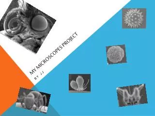
M Y Microscopes Project
M Y Microscopes Project. By JJ. The Light C ompound Microscope. In about 1597 two Dutch eyeglass makers, Zaccharias Janssen and his son

M Y Microscopes Project
E N D
Presentation Transcript
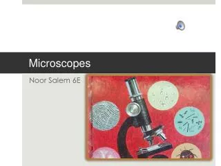
MY Microscopes Project By JJ
TheLight Compound Microscope In about 1597 two Dutch eyeglass makers, Zaccharias Janssen and his son Hans were looking at lenses in a tube. They saw that nearby objects looked at through two lenses in line were magnified. Their object was the first Compound Microscope. But, their lenses were pretty large and the magnification was only about 10X. Galileo also designed a Compound Microscope, but it was only useful for reflected light. Robert Hooke built the first useable British Compound Microscope in about 1655.
The Stereo Microscope The Stereo Microscope (or the dissecting microscope) has had a big impact on the medical and scientific communities. The man who invented the microscope was Horatio S. Greenough, an American instrument designer. The Stereo Microscope uses two different lenses to make different views for the left and right eye. This produces a three-dimensional view of the subject. Greenough took the plans for the Stereo Microscope to the Carl Zeiss Company of Jena in the early 1890s. Cherubind'Orleans produced the earliest version of a Stereo Microscope in 1671. While his model did have two lenses, d'Orleans' design made the left image to the right eyepiece and the right image to the left eyepiece, not making a true three-dimensional view. Horatio S. Greenough was the son of Horatio Greenough (1805-1852), a famous Boston sculptor. Horatio S. Greenough's middle name was Saltonstall. He shared the Saltonstall name with his uncle, Richard Saltonstall Greenough, who was also a sculptor like his brother.
The Transmission Electron Microscope (TEM) The transmission electron microscope was invented by two German scientists, Max Knoll and Ernst Ruska, in 1931. The transmission electron microscope (TEM) operates on the same basic principles as the light microscope but uses electrons instead of light. What you can see with a light microscope is limited by the wavelength of light. TEMs use electrons as "light source" and their much lower wavelength makes it possible to get a resolution a thousand times better than with a light microscope.
The Scanning electron Microscope (SEM) The scanning electron microscope (SEM) is a microscope that uses electrons instead of light to form an image. Since invented, in the early 1950's, scanning electron microscopes have made new areas of study in the medical and physical science communities. The SEM has allowed researchers to examine a much bigger variety of specimens. The idea of producing an image by scanning was first considered in 1842 when Alexander Bain invented the first fax machine. The idea of using charged particle beams (including electron beams) for scanning was later thought of in a German patent by Stintzing in 1929 (Stintzing H: Verfahrenund EinrichtungzumautomatischenNachweiss, Messung und Zählung von Einzellteilchenbeliebiger Art, Form und Grösse. The first scanned electron image was produced in 1935 by Max Knoll, co - inventor of the TEM, which received more attention at the time. It wasn't until 1952 that Sir Charles Oatley invented the SEM in its current form .
Bibliography http://en.wikipedia.org/wiki/Wiki http://static.arstechnica.net http://www.microscopesmall.com/ http://www.microscopeworld.com/MSWorld/images/ http://www.microscope-manufacturers.com/ http://www.aunet.com.au/imgs/microscopes/stereo_microscope/ http://www.uiowa.edu/~cmrf/methodology/tem/tem6.gif http://www.mauricewilkinscentre.org/bioviz/forstudents/ http://www.microscopehelp.com/images/06.jpg Thanks for watching