Peripheral
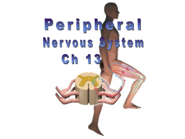
Peripheral. Nervous System. Ch 13. ____Cranial nerves attach to brain. ___Spinal nerves attach to spinal cord. 12 pair. 31 pair. Nerves. Bundles of axons in the PNS. Sensation: The conscious or subconscious awareness of external or internal stimuli. Perception:

Peripheral
E N D
Presentation Transcript
Peripheral Nervous System Ch 13
____Cranial nerves attach to brain ___Spinal nerves attach to spinal cord
12 pair 31 pair
Nerves Bundles of axons in the PNS
Sensation: • The conscious or subconscious awareness of external or internal stimuli. • Perception: • The conscious awareness and the interpretation of meaning of sensations.
Peripheral sensory receptors By location: • Exteroceptors • Sensitive to stimuli arising from outside body • Interoceptors • Or visceroreceptors, from internal viscera • Proprioceptors • Monitor degree of stretch in skeletal muscles, tendons, joints and ligaments
General Sensesvs. Special Senses • Pain • Temperature • Light touch • Pressure • Sense of body and limb position • Taste • Smell • Vision • Hearing • Balance
Sensory Receptors • Mechanoreceptors • Thermoreceptors • Photoreceptors • Chemoreceptors • Nociceptors • Osmoreceptors
General Senses Unencapsulated Nerve Endings Encapsulated Nerve Endings vs Free nerve endings skin, bones, internal organs, joints Deeper tissue, muscles Encapsulated nerve endings
Unencapsulated Nerve Endings • Free Nerve Endings • Pain & Temperature • Merkel’s Discs • Light Touch & Pressure • Root Hair Plexuses • Light Touch
Encapsulated Nerve Endings Meissner’s Corpuscles Discriminative Touch in Hairless Skin Areas Pacinian Corpuscles Deep Pressure Krause’s End-Bulbs Discriminative Touch in Mucous Membranes Ruffini’s Corpuscles Deep Pressure & Stretch (Proprioception)
The Epidermis Merkel cell Merkel Cells- slow mechanoreceptors (basal layer)
Skin Receptors free nerve endings Merkel disc Meissner’s corpuscles Ruffini corpuscle root hair plexus Pacinian corpuscles
Encapsulated Nerve Endings Proprioceptors Muscle Spindles - Skeletal Muscle Stretching Golgi Tendon Organs - Tendon Stretching Joint kinesthetic receptors – monitors stretch in synovial joints; sends info to cerebellum and spinal reflex arcs • to cerebrum, • cerebellum and • spinal reflex arcs
Muscle Spindle & Tendon Organ
Peripheral motor endings • Innervation of skeletal muscle • Innervation of visceral muscles and glands
Motor axons innervate skeletal muscle fibers at neuromuscular junctions = motor end plates
Motor unit: motor neuron & all the muscle fibers it innervates All muscles in motor unit contract together when neuron fires Stimulation of single motor unit causes weak contraction of entire muscle (spread out)
Innervation of visceral muscles & glands • Near end organ visceral motor axon swells = presynaptic terminals (vesicles with neurotransmitters): action slow (NT diffuses)
Pain- protective function Somatic Pain-results from injuries to skin, muscle, joints, tendon vs.Visceral Pain- pain in body organs
Nerve Damage & Repair in PNS • Mature neurons are amitotic • If the soma of a damaged nerve is intact, axon will regenerate • Involves coordinated activity among: • Macrophages—remove debris • Schwann cells—form regeneration tube and secrete growth factors • Axons—regenerate damaged part • CNS oligodendrocytes bear growth-inhibiting proteins that prevent CNS fiber regeneration
Endoneurium Schwann cells 1 The axon becomes fragmented at the injury site. Droplets of myelin Fragmented axon Site of nerve damage Figure 13.4 (1 of 4)
Macrophages clean out the dead axon distal to the injury. 2 Schwann cell Macrophage Figure 13.4 (2 of 4)
Axon sprouts, or filaments, grow through a regeneration tube formed by Schwann cells. 3 Aligning Schwann cells form regeneration tube Fine axon sprouts or filaments Figure 13.4 (3 of 4)
The axon regenerates and a new myelin sheath forms. 4 Site of new myelin sheath formation Schwann cell Single enlarging axon filament Figure 13.4 (4 of 4)
Cranial Nerves • On OldOlympus Towering Tops A Fat Voracious German Viewed A Hop • Olfactory- smell • Optic- vision • Oculomotor- 4 of the 6 extrinsic eye muscles • Trochlear- extrinsic eye muscles • Trigeminal- sensory fibers to the face and motor fibers to the chewing muscles • Abducens- controls eye muscles that turn the eye laterally • Facial- facial expression • Vestibulocochlear- hearing and balance • Glosopharyngeal- tongue and pharynx • Vagus- parasympathetic control of heart, lungs & abdominal organs • Accessory- accessory part of vagus nerve, neck & throat muscles • Hypoglossal- moves muscles under tongue
Olfactory Nerves I Olfactory bulb Olfactory tract Filaments of olfactory nerve Olfactory receptor cell
Abducens Nerves VI Lateral rectus muscle cut Abducens nerve
sensory pathway motor pathway Nerve Pathways into the Spinal Cord
Spinal nerves • Dorsal roots – sensory fibers arising from cell bodies in dorsal root ganglia • Ventral roots – motor fibers arising from anterior gray column of spinal cord Ventral root ganglia
Dorsal and ventral roots join in an intervertebral foramen forming spinal nerve • Outside foramen, re-branch as rami (sing., ramus):Dorsal and ventral rami (somatic) Rami communicantes (visceral) Spinal nerve
Dorsal rami serve the muscles and skin of the posterior trunk • Back, from neck to sacrum, innervated in a neatly segmented pattern: horizontal strip at same level as emergence from spinal cord • Ventral rami serve the muscles and skin of the lateral and anterior trunk • In thorax only, a simple segmented pattern as intercostal nerves • Also serve the limbs
Dorsal ramus Cross section of thorax showing main roots and branches of a spinal nerve • In the thorax, each ventral ramus continues as an intercostal nerve Ventral ramus Intercostal nerve
Spinal Nerves Nerve plexuses Cervical nerves C1-C8 • Networks of successive ventral rami that exchange fibers (crisscross & redistribute) • Mainly innervate the limbs • Thoracic ventral rami do not form nerve plexuses Thoracic nerves T1-T12 Lumbar nerves L1-L5 Sacral nerves S1-S5 Coccygeal nerve C0
Cervical Plexus • Formed by ventral rami of C1–C4 • Innervates skin and muscles of the neck, ear, back of head, and shoulders • Phrenic nerve • Major motor and sensory nerve of the diaphragm (receives fibers from C3–C5)