Download
1 / 1
10 likes | 131 Views
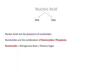
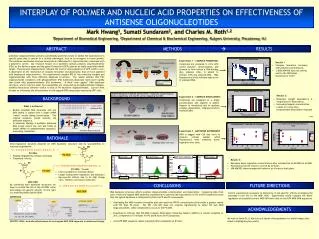
CHO blank. pd1EGFP blank. GFP F (FL1). GFP F (FL1). INTERPLAY OF POLYMER AND NUCLEIC ACID PROPERTIES ON EFFECTIVENESS OF ANTISENSE OLIGONUCLEOTIDES. Mark Hwang 1 , Sumati Sundaram 2 , and Charles M. Roth 1,2.

E N D
CHO blank pd1EGFP blank GFP F (FL1) GFP F (FL1) INTERPLAY OF POLYMER AND NUCLEIC ACID PROPERTIES ON EFFECTIVENESS OF ANTISENSE OLIGONUCLEOTIDES Mark Hwang1, Sumati Sundaram2, and Charles M. Roth1,2 1Department of Biomedical Engineering, 2Department of Chemical & Biochemical Engineering, Rutgers University, Piscataway, NJ ABSTRACT METHODS RESULTS Antisense oligonucleotides present a potentially powerful means to inhibit the expression of a specific gene that is central to a cellular phenotype, such as an oncogene in tumor growth. The antisense mechanism involved comprises an mRNA-specific oligonucleotide complexed with a polymeric carrier. Our research focuses on a synthetic cationic polymer, polyethylenimine (PEI), as the delivery agent and the green fluorescent (GFP) gene as an easily quantified model target. Fluorescence measurements indicate the level of downregulation of GFP, which in turn is dependent on the mechanism of complex formation and dissociation that we have assessed with biophysical measurements. Our experiments compare PEI of five molecular weights and oligonucleotides with three different backbone structures. The results indicate that PEI- oligonucleotide complexes with phosphodiester (PO) backbones dissociate much more rapidly than those with phosphorothioate (PS) backbones. A third “end capped” ODN backbone structure, where only the outer two nucleotides are connected by phosphorothioate linkages, exhibits dissociation behavior similar to that of PO backbone oligonucleotides. Current work focuses on evaluating the effectiveness of end-capped ODN using stably expressing GFP cells. Experiment 1 – COMPLEX FORMATION: Results 1 Complexes are prepared in vitro with varied polymer concentrations and allowed to form before a DNA binding dye, oligreen (OG), is added to interact with any unbound DNA. High fluorescence levels indicate high levels of unbound DNA. • Complex formation increases with polymer concentration. • 1.2KDa MW PEI does not bind as well to the ODN (high fluorescence level) PEI MW PEI MW HEPARIN MEDIATED COMPLEX DISSOCIATION - negatively charged heparin binds competitively with the PEI carrier, displacing the ODN. Results 2: Experiment 2 – COMPLEX DISSOCIATION: BACKGROUND PEI MW PEI MW • Molecular weight dependency is insignificant in dissociation. • Increasing heparin concentration results in a more than proportionate dissociation response Complexes are prepared at a single concentration and oligreen is added. Heparin in introduced last to mediate complex dissociation. Oligreen binds to released ODN. What is antisense? • Double stranded DNA separates and one DNA strand is copied onto a single mRNA “sense” strand during transcription. The original template strand encodes the antisense sequence. • In antisense therapy a synthetic antisense DNA strand enters the cell and binds to target mRNA of complementary sequence, preventing translation. PEI MW Experiment 3 - ANTISENSE EXPERIMENT: PEI MW PEI MW ODN is tagged with Cy5 dye (red) to measure cellular uptake, while fluorescence level measures down-regulation over time. RATIONALE Down-regulation duration depends on ODN backbone structure and its susceptibility to nuclease degradation. 5’…-A-G-G-U-C-A-C-U-U-U-G-C-A-A-C-G-…3’ • PO ODN • Rapidly degraded by cellular nucleases • Transitory effects. PO ODN GFP down-regulation ODN release Results 3: • Maximum down-regulation occurs 8 hours after introduction of AS ODN for all MW. • Fluorescence levels return to normal by 30 hours. • 10K MW PEI shows unexpected behavior at 4 hours in both plots. 5’…*A*G*G*U*C*A*C*U*U*U*G*C*A*A*C*G*…3’ • PS ODN ( * bonds) • Less susceptible to nuclease attack • Longer lasting down-regulation, but displays a • Non-specific effects due to its high charge ratio. Binds to non-target molecules. PS ODN GFP F (FL1) Cy5 F (FL4) MOD ODN By combining both backbone structures we hope to extend the life of the AS ODN, while decreasing non-specific effects. To the right is a modified (MOD) hybrid ODN: CONCLUSIONS FUTURE DIRECTIONS 5’…*A*G-G-U-C-A-C-U-U-U-G-C-A-A-C*G*…3’ MOD ODN DNA backbone structure affects polymer-oligonucleotide complexation and dissociation. Comparing data from each of the end-capped MOD sequence experiments to previous lab experiments on PO and PS sequences shows that the hybrid backbone is influenced by both its PO and PS components. Control experiments necessary to determine if non-specific effects accompany the extended in-cell life of the MOD ODN. Experiments would compare the down-regulation of scrambled control MOD ODN with that of anti-GFP MOD ODN sequences. MATERIALS • Overlaying the MOD complex formation plot onto previous PO/PS complexation plots yields a greater match with PO than PS plots. The PEI 1.2K MW does not complex significantly to either PO and MOD oligonucleotides, while complexation occurs in the PS ODN. • Experiments indicate that PEI-MOD complex dissociation following heparin addition is almost complete at 2hrs, compared to 15 minutes for PO and 8 hours for PS sequences. • Anti-GFP MOD sequence causes maximum down-regulation at 8 hours. ACKNOWLEDGEMENTS We wish to thank Dr. Li Kim Lee and Sandra Viriyayuthakorn for useful images that aided in highlighting key points. SPECIFIC GOAL: evaluate effectiveness of end-capped MOD ODN sequence in antisense therapy.