Download
1 / 54
540 likes | 860 Views
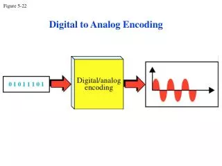
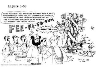
Figure 5-60. Page 122. EcoRI. Tetracycline Resistance. Ampicillin Resistance. PstI. HindIII. +. +. +. +. +. Origin of Replication. A plasmid. Figure 5-43 The pUC18 cloning vector. Page 106. Figure 5-46 Construction of a recombinant DNA molecule. Page 108.

E N D
Figure 5-60 Page 122
EcoRI Tetracycline Resistance Ampicillin Resistance PstI HindIII + + + + + Origin of Replication A plasmid
Figure 5-46 Construction of a recombinant DNA molecule. Page 108
Figure 31-1 The induction kinetics of b-galactosidase in E. coli. Page 1217
The lac operon • E-coli uses three enzymes to take up and metabolize lactose. • The genes that code for these three enzymes are clustered on a single operon – the lac Operon. What’s lactose??
Figure 31-2 Genetic map of the E. coli lac operon. Page 1218
What properties of macromolecules can we exploit to get them purified?
Figure 6-2 Solubilities of several proteins in ammonium sulfate solutions. Page 131
Figure 6-4 Solubility of b-lactoglobin as a function of pH at several NaCl concentrations. Page 132
Bacterial chlorophyll a Lamprey Hb
Table 6-1 Isoelectric Points of Several Common Proteins. Page 133
Sephadex: modified, cross-linked dextrans Fractionation Range (MW) G-50G-200Globular Proteins 1,500 - 30,000 5,000-800,000 Dextrans: 500 - 10,000 1,000-200,000 Sepharose 4B fractionation range 30,000-5,000,000 (Dextrans) 60,000-20,000,000 (Globular proteins)
Figure 6-9 Gel filtration chromatography. Porous beads allow small molecule entry but large molecules are excluded Page 137
Animation http://www.wiley.com/college/fob/quiz/quiz05/5-6.html
Absorbance Spectra for trp and tyr Phe absorbs a little as well. This phenomenon is the basis of one method to determine protein concentration in a non-destructive manner using Beer’s Law. Beer Beer Beer A = abc or bc
Figure 6-10 Molecular mass determination by gel filtration chromatography. Page 138
Ion Exchange Chromatography --among the most precise and frequently used methods for the fractionation of biological substances. Biomolecules with even small differences in charge can be separated. Very high resolution is obtained by choosing the optimal ion exchanger and the optimal separation conditions.
Excessively high salt concentrations cause shielding of the charges on the protein surface and effective binding to an exchanger can no longer take place. Since the bound biomolecules are subsequently displaced with the aid of an increasing salt gradient proteins varying in charge can be separated. The desorption of the proteins from the column begins only with increasing salt concentrations or pH changes, when the protein loses charge. The substances that have a higher charge density, are bound correspondingly stronger to the column while the others elute rapidly. The size of the sample volume in ion exchange chromatography is of secondary meaning as long as the initial solvent is of low eluting strength, so as not to allow any separation to occur. Under such conditions, the sample components are collected at the top of the column,
Some examples of exchanger functional groups Weak cation exchanger: carboxylic acid -CH2COO- Strong cation exchanger: sulfonate R-SO3- Weak anion exchanger: secondary or tertiary amine derivatives -CH2NHR2+ Strong anion exchanger: -CH2NR3 +
Functional Group pK Value Characteristic Description TMAE-Group > 13 strongly basic Trimethylammoniumethyl- DEAE-Group pK 11 weakly basic Diethylaminoethyl- DMAE-Group pK 8-9 weakly basic Dimethylaminoethyl- COO-Group pK 4.5 weakly acidic Carboxy- SO3-Group pK < 1 strongly acidic Sulfoisobutyl- SE-Group pK < 1 strongly acidic Sulfoethyl-
Common Buffers for IEC of proteins MoleculepKaCounter ion N-methyl piperazine 4.75 chloride piperazine 5.68 chloride or formate L-histidine 5.96 chloride bis-Tris 6.46 chloride bis-Tris propane 6.80 chloride triethanolamine 7.76 chloride or acetate Tris 8.06 chloride N-methyl-diethanolamine 8.52 chloride diethanolamine 8.88 chloride 1,3-diaminopropane 8.64 chloride ethanolamine 9.50 chloride piperazine 9.73 chloride 1,3-diaminopropane 10.47 chloride piperidine 11.12 chloride phosphate 12.33 chloride Which would you use for proteins?
Figure 6-11 Use of dialysis to separate small and large molecules. Page 139
Figure 6-15 Purification of staphylococcal nuclease by affinity chromatography. Page 141
Figure 6-20 Apparatus for slab gel electrophoresis. Page 146
Figure 6-23 Detection of proteins by immunoblotting. Page 148
Figure 6-25 Logarithmic relationship between the molecular mass of a protein and its relative electrophoretic mobility in SDS-PAGE. Page 149
Figure 6-27 Two-dimensional (2D) gel electrophoresis. Page 150