Microbiology
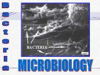
Microbiology. Clinical Pathology. Microbiology. The study of microscopic organisms Clinical microbiology is the identification of these organisms that cause clinical illness. Laboratory Safety. Most microorganisms are potentially pathogenic DO NOT EAT OR DRINK IN LAB!


Microbiology
E N D
Presentation Transcript
Microbiology Clinical Pathology
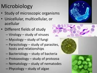
Microbiology • The study of microscopic organisms • Clinical microbiology is the identification of these organisms that cause clinical illness
Laboratory Safety • Most microorganisms are potentially pathogenic • DO NOT EAT OR DRINK IN LAB! • Clean area with disinfectant at beginning and end of the work period • Avoid putting any object in mouth (pencil, fingers). • Tape shut all containers of cultures before disposal. • Flame wire loops immediately after use. • Always use aseptic technique
Laboratory Safety Continued • Place contaminated materials in appropriate containers for disposal. • Never use a mouth pipette. • WASH HANDS • REPORT ALL LABORATORY INCIDENTS IMMEDIATELY!!!!
Equipment and Supplies • Incubator • Sterilizing heat source • Bunsen burner • Heating element • Alcohol lamps • Media • Microscope • Antimicrobrial sensitivity discs • Miscellaneous other equipment: • Metal loop • Slides • Sterile cotton tip applicators • Wax markers • Stain
Incubators • Allows organism to be grown under controlled conditions • Keeps specimen at 37 C (98.6 F) • Human body temperature and room air Oxygen • Most cultures are grown overnight and held at least 48 hours.
Sterilizing Heat Sources • Bunsen Burner • Sterilizes metal loop used for transferring organisms to be inoculated into growing media • Electric heating element • Usually ceramic, an enclosed heater. • Eliminates the need for a natural gas • Alcohol lamps • Less expensive • Glass lamp with wick • Does not sterilize metal loops that quickly
Media • Nutritive media- grows all types of bacteria • Selective media- grows only certain types of bacteria • Gram negative or gram positives • Enriched media- basic nutrient media with extra nutrients added-blood, serum, or egg. • Differential Media- contains elements that differentiate certain types of bacteria (ex. Lactose fermenters or hydrogen sulfide producers)
Media Continued • Examine media for accidental bacteria/fungal contaminants before use • Incubate al plates UPSIDE DOWN • Prevents condensation from dripping onto cultures. • Media may be solid (agar) or liquid (broth). • Plate is a flat, round container of agar • Tube is a screw-top container that may contain broth and agar • Slant, a tube of agar that has been allowed to gel at an angle.
Basic Nutrient Media • Peptone- hydrolyzed protein that can be metabolized by bacteria (provides AA) • Principal nutrient of the medium • Salt • Dextrose- gives carbon and energy for bacteria • Water • Meat extract- provides water soluble CHO nitrogen and vitamins • Solidifying agents- Agar, gelatin • Basic Nutrient Types: • Nutrient Agar • Trypticase soy agar (TSA)
Selective Media • Blood Agar • Chocolate Agar Media supports most bacterial pathogens
Blood Agar • TSA + 5% sheep blood • Can refer to Blood Agar Plate (BAP) • Blood agar should be bright red • Brownish-red color may indicate: • Blood is too old, RBC’s are hemolyzing • Blood was added to the agar base when the medium was too hot • Inadequate mixing of blood and agar
Blood Agar Continued • Also acts as a differential media • Four types of hemolysis: • Alpha hemolysis- partial hemolysis • Narrow band of greenish slimy discoloration around the colonies • Beta hemolysis- complete hemolysis • Clear zone around the bacterial colony • Gamma hemolysis- no hemolysis • Delta hemolysis- Double zone hemolysis • Double ring of hemolysis around colonies • Can differentiate different species of Streptococcus.
Chocolate Agar • For Hemophilus spp. • A very nutritive media • Hemolyzed RBC’s with growth factors • Has increased Carbon dioxide
MacConkey Media • Both a selective and differential media • Selects for Gram-negative • Uses crystal violet as a gram + inhibitor • Suppresses growth of gram + • Indicators • Lactose and neutral red • Lactose fermentors (E. coli, Enterobacter, and Klebsiella) produce acid from lactose and grow as pinkish-red colonies • Lactose non-fermentors- produce colorless colonies • Ex. No growth on MAC and good growth on BAP suggests Gram + • Inhibits swarming of Proteus
Enriched Media • Brain-heart Infusion Broth (BHIA) • Mueller-Hinton agar (MH) • For antibiotic sensitivity
Brain-heart Infusion Broth (BHIA) • General purpose broth used to increase the numbers of organisms before they are plated • Ex. Listeria from brain tissue may be difficult to grow, finely cut brain tissue is incubated for weeks in BHIA, then cultured.
Differential Media (several types) • Urea agar slant • Will turn from peach to pink with ammonium production • Triple sugar iron agar • Have lactose, sucrose, glucose • Differentiates- Salmonellas and other enteric bacteria • Mannitol salt agar • For select Staph species
Inoculating Culture Media • Use sterile technique • Flame neck of tube when transferring organisms. • Do not put the cap down but hold between last 2 fingers • Flame the near portion of the wire 1st, then work towards the contaminated end • Turns red • Prevents bacteria splatter
Streaking Plates • Flame the bacterial loop between and cool • Each streak is overlapped only 1-2 times to avoid depositing excessive numbers of bacteria • Will give discreet isolated colonies • Use the entire plate to streak • Turn plate for each streak • Flame/cool loop in between
Materials you will need: • Gloves • Inoculating loop OR sterilized wooden sticks for streaking • Permanent marker or grease pencil to label your plates beforehand • Bunsen burner or sterilizing heater if intending to sterilize the inoculating loop between streaks • Swab for collecting the primary inoculum, if intending to collect bacteria from an environmental source • Agar plate • Incubator, if incubating at a controlled temperature, such as 37。C. However, many common microbial species will grow on plates left at room temperature, through their growth may be slower than if the plates were incubated at 37。C • Antibacterial soap to wash your hands • 5% bleach solution to clean your work area when finished.
Step One: (The Primary Streak) If you are right-handed, hold the plate in your left hand, and the inoculating loop in your right - as through you would a paint brush. If you are left-handed, use the opposite hands. Touch your inoculating loop (sterile swab, or sterile stick as shown in the picture) to the material you want to spread. Go back and forth a number of times in a small area of the Agar plate. The goal is to spread your material completely over this inital area of the plate.
Step Two:(The Secondary Streak)Sterilize your inoculating loop, or use a fresh, sterile inoculating stick or swab. Make sure the loop is cool before your next streak. If you were to use the original loop, you will not be diluting the individual microbes you applied in the first streak. Pick up the plate and rotate it 1/4 of a turn to your left (if right-handed), or to your right (if left handed). Run the loop through the previous streak 2-3 times, then draw it along 1/3 of the remaining plate, as shown by the blue line in the image.
Step Three:(The Tertiary Streak)Rotate the plate another 1/4 turn and sterilize yourinoculating loop or take a fresh, sterile stick or swab. Again, make sure to cool your loop between streaks. Run the loop through the previous, secondary streak 2-3 times, and draw the streak over a remaining 1/3 of the plate, as shown.
Step Four:(The Quarternary Streak)Rotate the plate another 1/4 turn and sterilize the inoculating loop. Again, cool the loop between streaks, or use a new sterile swab. Run the loop through the previous tertiary streak 2 times and draw over the remaining free space in the plate, being careful not to contact the primary streak (yellow).
Inoculating Slants • Only the surface • Streak “S” shaped • Both surface and butt • Stab the butt • Withdraw the wire up same insertion path • Then streak the slant
Colony characteristics • Size- pin point, medium, large • Color- yellow, white, gray, cream, etc. • Density- opaque, transparent • Elevation-raised, flat, convex, droplike • Form-circular, irregular • Consistency- buttery, brittle, sticky • Odor- pungent, sweet, etc • Hemolysis- alpha, beta, gamma
Form What is the basic shape of the colony? For example, circular, filamentous, etc.
Elevation What is the cross sectional shape of the colony? Turn the Petri dish on end.
Margin What is the magnified shape of the edge of the colony?
Surface How does the surface of the colony appear? For example, smooth, glistening, rough, dull (opposite of glistening), rugose (wrinkled), etc.
Opacity For example, transparent (clear), opaque, translucent (almost clear, but distorted vision, like looking through frosted glass), iridescent (changing colors in reflected light), etc.
Chromogenesis (Pigmentation) For example, white, buff, red, purple, etc.
Bacterial Staining • Gram’s stain • Acid fast stain • Geimsa stain Examine number, types of bacteria
Gram Staining • Used to categorize bacteria as Gram + or Gram -. • Use cell wall morphology • Use young colonies (24 hours old) • Older colonies may not give proper results • Decolorization • Bacteria that retain the crystal violet-iodine complex stain Purple are Gram + • Those that lose the crystal violet and stain Red by safarin or basic fuschsin are Gram -
Gram Staining • Obtain a sample from one colony with sterile wire loop • Mix with drop of saline or water on the slide, if from broth use 2-3 loopfuls • Circle the area with wax pencil • After drying, heat fix- DO NOT overheat • Prevents bacteria from washing off • Kills bacteria and makes them pick up stain
Gram Staining • Exam also morphology • Bacilli • Cocci • Coccobacilli • Spiral • Paris, chains, clusters • Sometimes get a Gram variable reaction • Both Gram + and Gram – on same organism
Gram variable • May occur because: • Excessive heat fixation • Decolarization • Overly thick smear • Old cultures • Poor quality stain
Potassium Hydroxide Test (KOH) • Checks the gram reaction • Place a loopful of 3% KOH on slide • Place a large amount of surface growth • Stir, then slowly lift loop up after 30 seconds • Gram neg- sticky strand • Grand +- does not form a strand on lifting
Size/shape/arrangement • Cocci- spherical in shape • Staph aureus • Bacillus- rod in shape • Bacillus anthracis • Spiral- • Loose spirals- Borrelia • Tight spirals- Leptospira • Comma shaped spirals- Campylobacter
Bacterial arrangement • Single- most bacilli • Pairs- Streptococcus pneumoniae • Clusters- Staph aureus • Chains- Strep species • Palisades- Chinese letter pattern- Corynebacteria
Acid Fast stain • Several types of stains • Used to detect Mycobacteria and Nocardia • Acid fast bacteria- stain red • Non acid fast- stain blue or green • Depends on stain used- brilliant green or methylene blue
Giemsa stain • Used to detect spirochetes and ricketssiae • Demonstrates the capsule of Bacillus anthracis
Procedure for Identifying Bacteria • Streak on Blood agar and MAC • Incubated 18-24 hours • Select colonies from BAP vs. MAC • MAC may inhibit some colonies • Gram stain • Differential media
Gram Positive Cocci • Staphylococci • Steptococci • Micrococci
Catalase Test • Do when have a gram positive cocci and small gram positive bacilli (any gram positive colony) • Tests for the enzyme catalase, which acts on hydrogen peroxide to produce water and oxygen. • Place a small amount of an isolated colony from a blood agar plate on slide and a drop of catalase reagent (3% hydrogen peroxide). • Catalase positive- gas bubbles are produced • Catalase negative- no gas bubbles are produced • Do not transfer any blood agar with colony because may get a slightly positive reaction