Download
1 / 34
560 likes | 2.4k Views
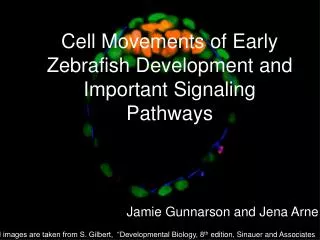
Cell Movements of Early Zebrafish Development and Important Signaling Pathways. Jamie Gunnarson and Jena Arne. All images are taken from S. Gilbert, “Developmental Biology, 8 th edition, Sinauer and Associates. Embryogenesis. Early development of

E N D
Cell Movements of Early Zebrafish Development and Important Signaling Pathways Jamie Gunnarson and Jena Arne All images are taken from S. Gilbert, “Developmental Biology, 8th edition, Sinauer and Associates
Embryogenesis Early development of zebrafish from the one-cell zygote to vertebrate embryo
Fertilization Activated zebrafish embryo. Arrow indicates point of sperm entry. Factors, such as beta-catenin mRNA needed for dorsal-ventral axis formation, are loaded into the egg. 200 μm Poleo G A et al. Biol Reprod 2001;65:961-966 ©2001 by Society for the Study of Reproduction
Cleavage Stage The fertilized egg undergoes synchronous cleavage. This egg is telolecithal, meaning only a small region is free of yolk. This yolk-free region is termed the blastodisc and divides by meroblastic, discoidal cleavage.
Blastula StageMid-Blastula Transition Blastula stages begin at the eighth division (128 cell stage). The pulsing characteristic of synchronous division stops and asynchronous division begins. At this point transcription of the zygotic genome begins. Between the ninth and tenth division, the Yolk Syncytial Layer (YSL) is formed by the fusion of cells with yolk.
Blastula Stagecontinued The blastomeres begin to mix randomly and mixing continues through late gastrulation. Dorsal YSL begins to express the nodal signal Squint when nuclear beta-catenin accumulates.
Blastula StageFate map The mixing of the cells finally allows for fate mapping during late-blastula. Text
Animal Pole Nodal Ventral Dorsal Vegetal Pole Blastula StageLate Blastula All embryonic cells undergo epiboly driven by the expansion of the YSL. The outermost layer of blastomeres (the enveloping layer) surrounds the yolk cell. Marginal blastomeres express Znr proteins (squint and cyclops) creating a nodal gradient causing different cell fate specification. Cells with very high nodal become prechordal plate, while cells with decreased nodal become notochord.
GastrulationEmbryonic Shield Formation Cells undergo dorsal convergence to form the embryonic shield. This structure organizes gastrulation. The embryonic shield secretes nodal which sets up the dorsal-ventral gradient, in which high nodal produces dorsal structures and low nodal produces ventral structures.
GastrulationMesendoderm Formation Cells of the embryonic margin begin to involute, giving rise to the mesendoderm. These specified cells involute to form the inner cell layer (hypoblast). Cells of the embryonic margin begin to involute, giving rise to the mesendoderm. These specified cells involute to form the inner cell layer (hypoblast).
Animal Pole BMP Ventral Dorsal Vegetal Pole GastrulationD/V Axis Formation by BMP Inhibitors BMP is high on the ventral side and turned off on the dorsal side by the BMP inhibitors Noggin, Chordin, and Follistatin. Inhibitors are expressed from the embryonic shield, and cell fates are specified along the dorsal-ventral axis.
Gastrulation Anterior-Posterior Axis Formation The anterior-posterior axis is formed by involution. The first cells to involute make anterior structures and the last cells to involute make posterior structures.
Gastrulation Convergent Extension Mesendoderm and ectoderm converge to the dorsal side and extend along the anterior-posterior axis. Convergent extension, epiboly and involution occur at the same time. Courtesy of Dr. Jennifer Liang Spring 2010
Question 1 • What is the importance of loading beta-catenin mRNA into the Zebrafish embryo?
Answer • Beta-catenin mRNA is necessary for dorsal-ventral axis formation.
Question 2 • What type of cleavage does the Zebrafish embryo undergo?
Answer • The zebrafish undergoes synchronous, meroblastic, discoidal cleavage.
Question 3 • What do YSL and MBT stand for and how are they related?
Answer • YSL stands for Yolk Syncytial Layer and MBT stands for Mid-Blastula Transition. The YSL forms during MBT.
Question 4 • Why is the mixing of the blastomeres important?
Answer • Without the mixing of blastomeres, fate mapping cannot take place.
Question 5 • What is epiboly driven by?
Answer • Epiboly is driven by the expansion of the YSL and causes the enveloping layer to surround the yolk.
Question 6 • Which structure secretes nodal and how does this signal affect dorsal-ventral axis formation?
Answer • The embyronic shield secretes nodal. Regions with high nodal produce dorsal structures and regions with low nodal produce ventral structures.
Question 7 • Which cells involute to form the inner cell layer (hypoblast)?
Answer • Mesoderm and endoderm, termed mesendoderm cells, involute to form the inner cell layer and are derived from the embryonic margin.
Question 8 • Explain the role of BMP inhibitors in dorsal-ventral axis formation.
Answer • The role of BMP inhibitors in dorsal-ventral axis formation is to turn off BMP signaling. This occurs on the dorsal side, thus causing BMP to accumulate on the ventral side, forming ventral structures.
Question 9 • Which cell movement is responsible for anterior-posterior axis formation? Explain how this occurs.
Answer • Involution is responsible for anterior-posterior axis formation. The first cells to involute give rise to anterior structures and the last cells to involute give rise to posterior structures.
Question 10 • What occurs during convergent extension?
Answer • During convergent extension all germ layers converge to the dorsal side of the developing embryo and extend along the anterior-posterior axis.