Download
1 / 77
770 likes | 791 Views
Explore the vertebrate endocrine system, hormone actions, and hormonal balance in the body for optimal health. Learn about the roles of different glands and hormone-secreting sources.

E N D
Endocrine Control Chapter 35
Impacts, IssuesHormones in Balance • Many chemicals we release into the environment (such as the herbicide atrazine) have disruptive hormonal effects
35.1 Introducing the Vertebrate Endocrine System • Animal cells communicate with one another by way of a variety of short-range and long-range chemical signals • Animal cells communicate with adjacent cells through gap junctions and by releasing molecules that bind to receptors in or on other cells
Shot-Distance Signaling • Neurotransmitters secreted by neurons diffuse across the synaptic cleft to the target cell • Local signaling molecules, such as prostaglandins released by injured cells, affect only neighboring cells
Long-Distance Signaling • Animal hormones secreted into interstitial fluid enter capillaries, are distributed throughout the body, and have wide-reaching effects • Pheromones diffuse through water or air and bind to target cells in other individuals (help integrate social behavior)
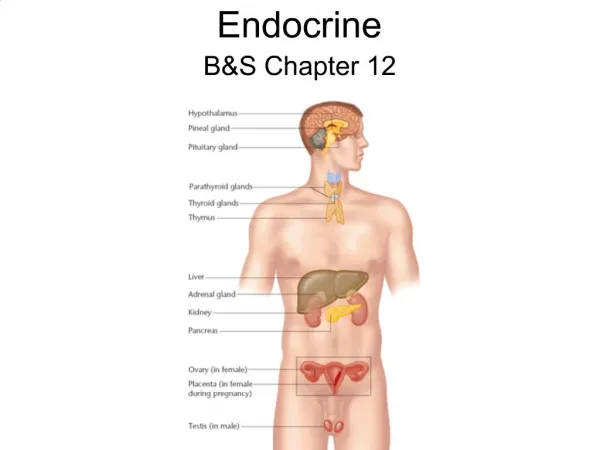
Overview of the Endocrine System • Hormones • Internal secretions carried by the blood that influence the activities of specific body organs • Endocrine system • Glands and other hormone-secreting sources
Nervous-Endocrine Interactions • Nervous and endocrine systems interact • Both respond to the hypothalamus, a command center in the forebrain • Most organs receive and respond to both nervous signals and hormones
hypothalamus closer view of the hypothalamus and pituitary gland Hypothalamus Makes and secretes releasers and inhibitors, hormones that act in the anterior lobe of the pituitary. pituitary gland Pituitary gland Pineal gland Anterior lobe makes and secretes ACTH, TSH, LH, FSH (stimulate secre- tion by other endocrine glands), pro- lactin (acts on mammary glands) and growth hormone (affects overall growth). Makes and secretes melatonin (affects sleep/wake cycles, onset of puberty). Thyroid gland Makes and secretes thyroid hormone (metabolic and developmental effects) and calcitonin (lowers blood calcium). Adrenal glands (one pair) Adrenal cortex makes and secretes cortisol (affects metabolism, immune response), aldosterone (acts in kid- neys), small amount of sex hormones. Parathyroid glands (four) Make and secrete parathyroid hormone (raises blood calcium level). Ovaries (one pair of female gonads) Thymus gland Make and secrete progesterone and estrogens (affect primary sex organs and influence secondary sexual traits). Makes and secretes thymosins (act in maturation of T cells, a type of white blood cell). Pancreas Testes (one pair of male gonads) Makes and secretes insulin (lowers blood glucose level) and glucagon (raises blood glucose level). Make and secrete testosterone and other androgens (affect primary sex organs and influence secondary sexual traits). The Human Endocrine System Fig. 35-2, p. 599
35.2 The Nature of Hormone Action • Cell communication involves three steps • For a hormone to have an effect, it must bind to protein receptors on or inside a target cell
Intracellular Receptors • Steroid hormones are made from cholesterol and can diffuse across the plasma membrane • Most steroid hormones form a hormone-receptor complex that binds to a promoter inside the nucleus and alters the expression of specific genes
Receptors at the Plasma Membrane • Large amine, peptide and protein hormones bind to a receptor at the plasma membrane • Binding triggers formation of a second messenger (molecule that relays signal into cell) • Enzyme converts ATP to cAMP • cAMP activates a cascading series of reactions
Hormone Actions Step 1 A steroid hormone molecule is moved from blood into interstitial fluid bathing a target cell. Step 1 A peptide hormone molecule, glucagon, diffuses from blood into interstitial fluid bathing the plasma membrane of a liver cell. unoccupied glucagon receptor at target cell’s plasma membrane Step 3 The hormone diffuses through the cytoplasm and nuclear envelope. It binds with its receptor in the nucleus. cyclic AMP + Pi ATP Step 2 Glucagon binds with a receptor. Binding activates an enzyme that catalyzes the formation of cyclic AMP from ATP inside the cell. Step 5 The resulting mRNA moves into the cytoplasm and is transcribed into a protein. receptor hormone– receptor complex gene product Step 2 Being lipid soluble, the hormone easily diffuses across the cell’s plasma membrane. Step 3 Cyclic AMP activates another enzyme in the cell. Step 4 The hormone– receptor complex triggers transcription of a specific gene. Step 4 The enzyme activated by cyclic AMP activates another enzyme, which in turn activates another kind that catalyzes the break- down of glycogen to its glucose monomers. Step 5 The enzyme activated by cyclic AMP also inhibits glycoge synthesis Stepped Art Fig. 35-3, p. 601
Hypothalamus GnRH Anterior Pituitary FSH, LH Gonads Sex hormones Generalized diagram showing control of sex hormone secretion. Stepped Art Fig. 35-17, p. 612
Receptor Function and Diversity • Only cells with appropriate and functional receptor proteins can respond to a hormone • Gene mutations that alter receptor structure can prevent or change cell response to a hormone • Examples: Androgen insensitivity syndrome, variations in ADH receptors
35.1-35.2 Key ConceptsSignaling Mechanisms • Hormones and other signaling molecules function in communication among body cells • A hormone travels through the blood and acts on any cell that has receptors for it • The receptor may be at a target cell’s surface or inside the cell
35.3 The Hypothalamus and Pituitary Gland hypothalamus anterior lobe of pituitary posterior lobe of pituitary Fig. 35-4, p. 602
Hypothalamus • Hypothalamus • Main center for control of internal environment • Lies deep inside the forebrain and interacts, structurally and functionally, with the pituitary gland
Pituitary Gland • Pituitary gland • Posterior lobe secretes hormones made in the hypothalamus • Anterior lobe synthesizes its own hormones • The hypothalamus signals the pituitary by way of secretory neurons that make hormones
Posterior Pituitary Function • Some secretory neurons of the hypothalamus make hormones that move through axons into the posterior pituitary, which releases them • Antidiuretic hormone (ADH) • Oxytocin (OT)
A Cell bodies of secretory neurons in hypothalamus synthesize ADH or oxytocin. B The ADH or oxytocin moves downward inside the axons of the secretory neurons and accumulates in the axon terminals. C Action potentials trigger the release of these hormones, which enter blood capillaries in the posterior lobe of the pituitary. Interactions of Hypothalamus and Posterior Pituitary D Blood vessels carry hormones to the general circulation. Stepped Art Fig. 35-5, p. 603
Anterior Pituitary Function • Other hypothalamus neurons produce releasers and inhibitors carried by blood that regulate secretion of anterior pituitary hormones • Adrenocorticotropic hormone (ACTH) • Thyroid-stimulating hormone (TSH) • Follicle stimulating hormone (FSH) • Luteinizing hormone (LH) • Prolactin (PRL) • Growth hormone (GH)
A Cell bodies of secretory neurons in hypothalamus synthesize inhibitors or releasers that are secreted into the stalk that connects to the pituitary. B The inhibitors or releasers picked up by capillaries in the stalk get carried in blood to the anterior pituitary. C The inhibitors or releasers diffuse out of capillaries in the anterior pituitary and bind to their target cells. Interactions of Hypothalamus and Anterior Pituitary D When encouraged by a releaser, anterior pituitary cells secrete hormone that enters blood vessels that lead into the general circulation. Stepped Art Fig. 35-6, p. 603
Feedback Controls of Hormone Secretion • Positive feedback mechanisms • Response increases the intensity of the stimulus • Example: Oxytocin and childbirth contractions • Negative feedback mechanisms • Response decreases the stimulus
35.4 Growth Hormone Function and Disorders • Excessive growth hormone (GH) causes faster than normal bone growth • Occurrence in childhood results in gigantism • Occurrence in adulthood results in acromegaly • A deficiency of GH during childhood can cause dwarfism
35.3-35.4 Key ConceptsA Master Integrating Center • In vertebrates, the hypothalamus and pituitary gland are connected structurally and functionally • Together, they coordinate activities of many other glands • Pituitary hormones affect growth, reproductive functions, and composition of extracellular fluid
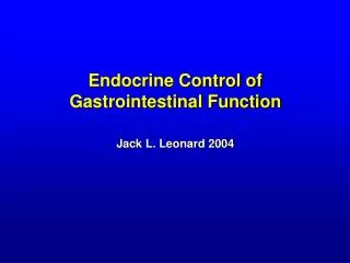
35.5 Sources and Effects of Other Vertebrate Hormones • In addition to the hypothalamus and pituitary gland, endocrine glands and endocrine cells secrete hormones • The gut, kidneys, and heart are among the organs that are not glands, but include hormone-secreting cells
Multiple Hormone Receptors • Most cells have receptors for multiple hormones, and the effect of one hormone can be enhanced or opposed by another one • Example: Skeletal muscle hormone receptors • Glucagon, insulin, cortisol, epinephrine, estrogen testosterone, growth hormone, somatostatin, thyroid hormone and others
35.6 Thyroid and Parathyroid Glands • The thyroid regulates metabolic rate, and the adjacent parathyroids regulate calcium levels
thyroid cartilage (Adam’s apple) Thyroid Gland trachea (windpipe) anterior Fig. 35-8a, p. 606
epiglottis pharynx Thyroid Gland Parathyroid Glands posterior Fig. 35-8b, p. 606
The Thyroid Gland • Thyroid gland • Located at the base of the neck; secretes iodine-containing thyroid hormones and calcitonin • Regulated by a negative feedback loop • Hypothyroidism • Low levels of thyroid hormone, caused by iodine deficiency or Graves’ disease, causes goiter
Negative Feedback Control of Thyroid RESPONSE STIMULUS + Hypothalamus Blood level of thyroid hormone falls below a set point. TRH Anterior Pituitary Rise of thyroid hormone level in blood inhibits the secretion of TRH and TSH. TSH Thyroid Gland Thyroid hormone is secreted. Stepped Art Fig. 35-9, p. 606
The Parathyroid Glands • Parathyroid glands • Release parathyroid hormone (PTH) in response to low blood calcium levels • Targets bone cells and kidney cells • Stimulates conversion of vitamin D to calcitriol
35.7 Twisted Tadpoles • Impaired thyroid function in frogs: An example of hormone-disruptor pollution in the environment • Includes pesticides, perchlorates
35.8 Pancreatic Hormones • Pancreas • Exocrine cells secrete digestive enzymes • Endocrine cells clustered in pancreatic islets stomach pancreas small intestine Above, location of the pancreas. Right, how cells that secrete insulin and glucagon react to shifts in the blood level of glucose. Insulin and glucagon work antagonistically to regulate glucose level, an example of homeostasis. (a) After a meal, glucose enters blood faster than cells can take it up. Its level in blood increases. (b, c) In the pancreas, the increase stops alpha cells from secreting glucagon and stimulates beta cells to secrete insulin. (d) In response to insulin, muscle and adipose cells take up and store glucose, and liver cells synthesize more glycogen. (e) The outcome? Insulin lowers the glucose blood level. (f) Between meals, the glucose level in blood declines. (g, h) This stimulates alpha cells to secrete glucagon and stops beta cells from secreting insulin. (i) In the liver, glucagon causes cells to break glycogen down into glucose, which enters the blood. (j) The outcome? Glucagon raises the amount of glucose in blood. Fig. 35-12 (top), p. 608
Insulin and Glucagon • Two pancreatic hormones with opposing effects work together to regulate blood sugar levels • Insulin • Increases cell uptake and storage of glucose • Secreted in response to high blood glucose • Glucagon • Increases breakdown of glycogen to glucose • Secreted in response to low blood glucose
Responses to Changes in Blood Glucose A Stimulus F Stimulus Increase in blood glucose Decrease in blood glucose Above, location of the pancreas. Right, how cells that secrete insulin and glucagon react to shifts in the blood level of glucose. Insulin and glucagon work antagonistically to regulate glucose level, an example of homeostasis. (a) After a meal, glucose enters blood faster than cells can take it up. Its level in blood increases. (b, c) In the pancreas, the increase stops alpha cells from secreting glucagon and stimulates beta cells to secrete insulin. (d) In response to insulin, muscle and adipose cells take up and store glucose, and liver cells synthesize more glycogen. (e) The outcome? Insulin lowers the glucose blood level. (f) Between meals, the glucose level in blood declines. (g, h) This stimulates alpha cells to secrete glucagon and stops beta cells from secreting insulin. (i) In the liver, glucagon causes cells to break glycogen down into glucose, which enters the blood. (j) The outcome? Glucagon raises the amount of glucose in blood. PANCREAS PANCREAS C beta cells H beta cells B alpha cells G alpha cells insulin glucagon glucagon insulin LIVER MUSCLE FAT CELLS LIVER D Body cells, especially those muscle and adipose tissue, take up and use more glucose. I Cells in liver break down glycogen faster. The released glucose monomers enter blood. Cells in skeletal muscle and liver store glucose in the form of glycogen. E Response J Response Increase in blood glucose Decrease in blood glucose Fig. 35-12 (right), p. 608
35.9 Blood Sugar Disorders • Glucose is the main energy source for brain cells and the only energy source for red blood cells • Having too much or too little glucose in blood causes problems throughout the body
Diabetes • Diabetes mellitus is a metabolic disorder in which cells do not take up glucose properly • Results in complications throughout the body • Type 1 diabetes (juvenile-onset diabetes) • Autoimmune disease that destroys insulin-producing cells; requires insulin injections • Type 2 diabetes (adult onset diabetes) • Target cells do not respond to insulin
Hypoglycemia • Hypoglycemia • Blood glucose levels low enough to disrupt normal body functions • Caused by excess insulin production or overdose of injected insulin in diabetics • Can cause dizziness, confusion, and shock
35.10 The Adrenal Glands • Adrenal glands • Sit atop kidneys • Have two parts (adrenal cortex and adrenal medulla) that are controlled by different mechanisms and release different hormones