Download
1 / 9
90 likes | 285 Views
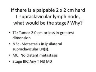
If there is a palpable 2 x 2 cm hard L supraclavicular lymph node, what would be the stage? Why?. T1: Tumor 2.0 cm or less in greatest dimension N3c -Metastasis in ipsilateral supraclavicular LN(s ). M0: No distant metastasis Stage IIIC Any T N3 M0. Differential diagnosis?.

E N D
If there is a palpable 2 x 2 cm hard L supraclavicular lymph node, what would be the stage? Why? • T1: Tumor 2.0 cm or less in greatest dimension • N3c -Metastasis in ipsilateral supraclavicular LN(s). • M0: No distant metastasis • Stage IIIC Any T N3 M0
Differential diagnosis? • Fibrocystic breast disease • Fibroadenoma • Carcinoma
fibrocystic breast disease • benign (noncancerous) condition characterized by round lumps that move freely within the breast tissue. This lumps are usually tender to the touch.
-Work-ups? Why? • 1. FNAB • 2. Mammography • 3. Chest x-ray • 4. Ultrasound • 5. CT scan • 6. Bone scan
FNAB • A fine needle aspiration (FNA) is a quick, simple procedure which is done in the outpatient clinic. Using a fine needle and syringe, the doctor takes a sample of cells from the breast lump and sends it to the laboratory to see if any cancer cells are present.
Mammography • The goal of mammography is the early detection of breast cancer, typically through detection of characteristic masses and/or microcalcifications.
Chest x-ray • Before treatment begins, a chest x-ray may be done to rule out metastasis of breast cancer to your lungs. • determine your lung and heart capacity. • check for the possibility of pneumonia or lung inflammation. • Because your immune system may be low, it is important to get help for any new conditions that develop. • After treatment, a follow-up chest x-ray is done to make sure your cancer has not spread to your lungs, or anywhere else in your chest.
Ultrasound • An ultrasound uses sound waves to build up a picture of the breast tissue. Ultrasound can tell whether a lump is solid (made of cells) or is a fluid-filled cyst. It can also often tell whether a solid lump is likely to be benign or malignant.