Download
1 / 50
500 likes | 753 Views
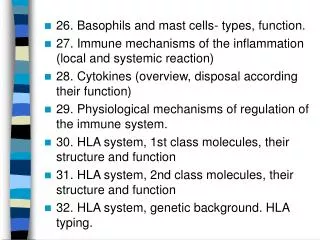
Basophils and mast cells, and their importance in immune responses. Mast cells

E N D
Basophils and mast cells, • and their importance in immune responses
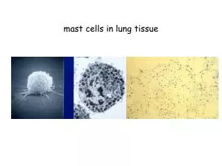
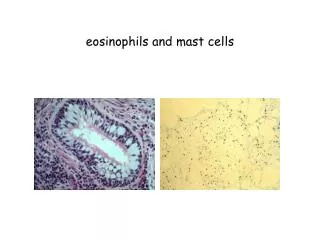
Mast cells • a) mucosal mast cells - location: in the mucous membrane of the respiratory and gastrointestinal tract - produce: histamine, heparin, serotonin, tryptasys, cytokines, and other enzymes, leukotriene C4 - participation: allergy and parasitosis, increased in the activation of TH2 b) connective tissue mast cells- localization: predominantly in the connective tissue - produce: tryptasys, chymasys and other enzymes, prostaglandin D2 - participation: the allergy and parasitosis are not participating, increased when fibrosis
Features mast cells- defense against parasitic infections - regulating the immune response - apply during inflammation, in angiogenesis, in tissue remodeling - participate in the maintenance of physiological functions of mucosal - contribute to the normal metabolism of connective tissue - communication between the immune and nervous system - the pathological circumstances are responsible for the early type hypersensitivity
Schema activation of mast cells - establish multivalent antigen (multicellular parasite) using the IgE to highaffinnity Fc receptor for IgE (FcRI) - aggregation of several molecules FcRI initiate mast cell degranulation (cytoplasmic granules merger with the surface membrane and release their contents) - activation of arachidonic acid metabolism (leukotriene C4, prostaglandin PGD2) - start of production of cytokines (TNF, TGF , IL-4, 5,6 ...)
Mast cell receptors- highaffinity Fc receptor for IgE (FcRI) - Fc receptor for IgG - receptor for complement fragment C5a (activated degranulation independent of IgE) - surface adhesion molecules ( integrins, L-selectin) - growth factor receptors (IL-3, IL-4, SCF)
Secretory products of mastcells- cytoplasmatic granules: hydrolytic enzymes, proteoglycans (heparin, chondroitin sulphate), biogenic amines (histamine, serotonin) Histamine causes vasodilation, increased vascular permeability, erythema, edema, itching, contraction of bronchial smooth muscle, increases intestinal peristalsis, increased mucus secretion mucosal glands in the respiratory tract and GIT. - arachidonic acid metabolites (leukotriene C4, prostaglandin PGD2) - cytokines (TNF, TGF IL-4, 5, 6 ...)
Basophils • - differentiate from myeloid precursor - are considered to be the circulating form of mast cells - receptor equipment, containing granules, the mechanisms of stimulation and functions are very similar to mast cells • - are responsible for the emergence of anaphylactic shock
Immune mechanisms of inflammation (Local and systemic reactions)
InflammationIs a summary of physiological responses to breach the integrity of the organism, leading to protection against infection damaged sites, localization of damage and healing. The first signals to the development of inflammatory responses come from mast cells, phagocytes, and the substances released from damaged cells and extracellular components of matter. Inflammation - acute (physiological defensive reactions, usually subside without consequences, damaged tissue heals completely) - chronic (usually already pathological reactions, occurs partial destruction of tissue and compensation with fibrous tissue) Response of the organism - local - system
Local body's response to inflammationManifestations - pain (dolor), heat (calor), redness (rubor), swelling (tumor) and loss of function (funkcio laesa) - increased permeability of blood vessels (vasoactive amines, complement components C3a, C5a, leukotrienes ..., swelling at site of inflammation) - increased expression of adhesion molecules on endothelia - activation of coagulation, fibrinolytic, kinin and complement system - influence of local nerve endings (prostaglandins, pain) - changes in temperature (IL-1, IL-6, TNF, prostaglandins)
Systemic response to inflammation-depends on the extent of damage and duration of local inflammation - fever (proinflammatory cytokines TNF, IL-1, IFN stimulate hypothalamic center of thermoregulation) - mobilization of tissue metabolism - induction of expression of Hsp (heat-shock-proteins; function as chaperones) - production proteins of acute phase (CRP, SAP, C4, C5; opsonization and complement activation)
- increased hepatic synthesis of certain serum transport proteins (ceruloplasmin, transferrin) • - increased synthesis of protease inhibitors ( macroglobulin) - leukocytosis Septic shock - the massive penetration of microorganisms into the bloodstream Anaphylactic shock - basophil degranulation and complement activation with allergen
Repair of damaged tissue- elimination of damaged cells with phagocytes - activation of fibroplastic mechanisms - activation of angiogenesis - regeneration and tissue remodeling
Cytokines - regulatory proteins and glycoproteins produced by leukocytes and other cells - essential regulators of the immune system - apply outside the immune system (angiogenesis, tissue regeneration, carcinogenesis, affecting a number of brain functions, embryonal development ...) cytokines - secreted - diaphragm (ensure local action, embedded in the membrane with sequences of about 20 hydrophobic AA; CD 80, CD86, CD40L, FasL)
- pleiotropic effect - operates in a cascade - cytokine network - cytokine system is redundant effects of cytokines - autocrine - paracrine - endocrine - are known as interleukins (exception: TNF, lymphotoxin, TGF, interferons, CSF and growth factors)
B cells communicate via cytokines with other inflammatory cells, such as T cells and macrophages
Overview of cytokines:- interleukins (IL-1 and IL-23) - chemokines (IL-8 and related molecules) - interferons (IFN---) - transforming growth factors (TGF TGF) - colony stimulating factors (G-CSF, M-CSF, GM-CSF) - tumors necrosis factors (TNF-, lymphotoxin) - other growth factors (SCF, EPO, FGF, NGF, LIF)
Distribution of cytokines by function:- proinflammatory cytokines (IL-1and , IL-6, 8, 12, 18, TNF) - antiinflammatory cytokines (IL-1Ra, IL-4, IL-10, TGF) - cytokines with the activity of hematopoietic cells growth factor (IL-2, 3, 4, 5, 6, 7, 9, 11, 14, 15, CSF, SCF, LIF, EPO) - cytokines applying in TH2 humoral immunity (IL-4, 5, 9, 13) - cytokines applying in the cell-mediated immunity TH1 (IL-2, 12, IFN, GM-CSF, lymphotoxin) - cytokines with anti-virus effect (IFN-, IFN-, IFN- )
1) The regulation with antigen- induction and extinction of the immune response - affinity maturation of B lymphocytes - maintenance of immunological memory - antigenic competing - the threshold density of complex MHC gpII-Ag on APC
2) The regulation with antagonistic peptidesagonist - antigenic peptide, which initiate a full answer of T cells (proliferation, differentiation TH or TC and stimulation of effector functions) • antagonist - (partial agonist) similar peptide structura to antigenic peptide, which causes qualitatively different T lymphocyte response (eg production of only some cytokines, anergy ...) • - negative signals induced by antagonist may overcome positive signals induced by agonist, which is in the body in excess - some micro-organisms inhibits reactivity of T cells with production mutant forms of their proteins, giving rise to antagonistic peptides - the possibility of therapeutic applications (transplantation, autoimmunity, chronic inflammation)
3) The regulation with antibody- antibody competes with BCR for the antigen (negativ regulator of B lymphocyte stimulating) - immunocomplexesIgGbind to B lymphocytes to BCR and FcR, the blocking effect of activation of B lymphocytes - immunocomplexes with C3dg have costimulating effects (C3dg binds to CR2 on the surface of B lymphocytes) - yet it is unclear meaning of regulation by idiotyp network
4) The regulation with cytokines and cellular contact • 4a) The interaction of APC - T lymphocyteTH precursor, which recognizes an infected macrophage and receive signals through the TCR, CD 28 and receptor for IL-12 and other adhesion and signaling molecules, proliferates and differentiates to the effector TH1 cells that produce IFN and IL-2. IFN promotes the conversion of macrophages to activated that produce NO (destruction of intracellular parasites) and cytokines IL-1, TNF ... (stimulation of local inflammation). IL-2 is the autocrine growth factor for TH1 cells. TH precursor, which recognizes an infected macrophage and receive signals through the TCR, CD 28 and receptor for IL-4 and receptor for IL-2 and other adhesion and signaling molecules, proliferates and differentiates to effector TH2 cells, which provide auxiliary signals to B lymphocytes using secreted cytokines IL-4, IL-5, IL-6 and through adhesion molecules CD 40L (binds to the costimulating receptor of B lymphocytes CD 40).
4b) Mutual regulation of activities TH1versus TH2 Whether the precursors TH lymphocytes will evolve in TH1 or TH2 decides ratio of cytokine IL-12 and IL-4. - IL-12 is produced by macrophages and dendritic cells stimulated by certain microorganisms - IL-4 is produced by basophils and mast cells - cytokines produced by TH1 (mainly IFN) inhibit the development of TH2 and stimulate the development of TH1 (IL-2 also stimulates TH2) - cytokines produced by TH2 (IL-4, IL-10) inhibit the development of TH1 and stimulate TH2 development - (development of TH3 is stimulated by specific cytokine environment IL-4, IL-10, TGFTH3 produce TGF and cooperate with B lymphocytes in MALT)
4c) Cytotoxic T lymphocytesTC precursor, which recognizes a complex of MHCgpI - antigenic peptide on the surface of APC via TCR and receives signals via CD 28, proliferates and differentiates to a clone of mature effector cytotoxic cells with the help of TH1 cells producing IL-2. Professional APC are dendritic cells or macrophages, which are infected with a virus or absorbed antigens from dead virus infected, tumor or stressed cells. In order the APC could activate precursor TC, must themselves be stimulated by contact with TH cells via CD 40, then dendritic cell begins express CD 80, CD86 and produce cytokines (IL-1, IL-12). Transformation of resting APC to activated. • 4d) NK cellsNK cells are capable without prior stimulation, proliferation and differentiation to kill tumor or virus-infected cells. Have on their surface stimulation (Fc receptor CD16) and inhibitory receptors (imunoglobulins, C lektins), the resulting response depends on what signals prevail.
4e) Extracellular contact - inhibitory receptors, negative regulation CTLA-4 (CD152) - ligand CD80, CD86 (activated T lymphocyte provides a strong inhibitory signal dampening excessive reactivity) KIR, CD94/NKG2 - Inhibitory receptors on NK cells Fas - Fas L (self-destruct interaction on the surface of activated effector T lymphocytes)
5) Suppression mediated by T lymphocytes- mutual negative interactions TH1a TH2 cytokine-mediated (TH2 lymphocytes produce IL-4 and IL-10 that suppress the immune response based on TH1 cells) - CD 8+ TS - suppressor T lymphocytes has not yet been isolated as a separate subpopulation(probably partly identical with TC) - negatively regulate the activation of other T cells (TH, TC) - liquidation of APC and TH with specific TC - creating cytokine environment favoring developmentofTH2 (some TCCD 8+ subpopulations produce IL-4, IL-10) - TS can act as anergic cells, which competes with other cells for the stimulating IL-2
- soluble suppressor factors - some CD 8+ T lymphocytes produce a soluble form of TCR • - clonal elimination or anergisation of T lymphocytes after contact with antigen on the surface of cells other than APC (missing costimulating signals), clonal elimination is important in establishing tolerance to autoantigens - regulatory T lymphocytes (CD 4+ TR1, similar TH3), which produce IL-10, help maintain tolerance to autoantigens
6) Neuroendocrine regulation • - some neurotransmitters act on leukocytes (noradrenaline) - direct contact free nerve endings with mast cells (pain when • mast cell degranulate) - some endocrine hormones act on leukocytes (steroids, growth hormone, thyroxine, endorphins ...) - leukocytes produce a number of hormones (endorphins, ACTH, TSH, growth hormone ...) - some cytokines act on the nervous system (IL-1, IL-6, LIF, TNF) - the effect of emotional stress on the immune system
7) Factors affecting the outcome of the immune response • The same antigen can induce an active immune response or an active state of tolerance, the result of response depends on many factors: - state of the immune system - characteristics of antigen - dose of antigen - method of administration
MHC glycoproteins class I • (major histocompatibility complex) - function of MHC gp.I is the presentation of peptide fragments that are produced by the cell,on the cell surface so as to be recognized by T lymphocytes (cytotoxic) - present in all cells of the organism - 3 isotypes of classical human MHC gp. (HLA - A,-B,-C) - 3 isotypes of non-classical MHC gp. (HLA - E,-F,-G; molecule CD1)
Structure of MHC gp. I- MHC gp. class I consists of transmembrane chain (45kDa) and non-covalently associated 2mikrotubulin (12 kDa) - chain has 3 domains, 2 N-terminal (1, 2 - binding site for peptides) and 1 C-terminal domain (3 - enshrined in the cytoplasmic membrane, a structure similar imunoglobulin domain) - binding site for the peptide is structurally eminent groove whose bottom is made up of structure and sides are bounded by 2 helixes - binding of peptide is necessary for a stable conformation of MHC gp, • thus ensure its long presentation on the cell surface
Binding of peptides • - MHC gp I bind peptides with a length of 8 to 10 aminoacides - certain molecule of MHC gp. bounds peptides sharing common structural features - coupling motif - important are aminoacides near the ends of peptide - the binding of endogenous peptides occurs in the endoplasmic reticulum during biosynthesis of MHC gp - after a string and 2mikrotubulin create in the ER folding into the correct conformation and the mutual association and the association with an appropriate peptide, the complex is further processed in the Golgi apparatus and then is presented on the cell surface - linked peptides derived from proteins degraded with proteasome, which cleaves cytoplasmic proteins determined for destruction (labeled with ubiquitin), peptide fragments are transported into the ER by specific membrane pump
Non-classical MHC gp I • - HLA - E,-F,-G; CD1 molecules - structurally similar to classical MHC gp - are less polymorphic - there are only on some cells - specialized for binding specific ligands
HLA - E - found on many cell types - binds signal (N-terminal) peptides from precursors of HLA - A,-B,-C,-G HLA - G - occurs on the trophoblast cells - binds many different peptides Complexes of HLA - E and HLA - G with peptides are recognized with inhibition receptors of NK cells and contribute to the tolerance of the fetus in utero. CD1 molecules - bind glycolipid or other highly hydrophobic substances (microbial or their body), these complexes are recognized by specialized T lymphocytes. • Others - like MHC gp I, but without the ability to bind peptides or other antigenic fragments - FcRn, MICA, MICB, HFE
MHC glycoproteins class II- function of MHC gp II is a presentation of peptide fragments from cell absorbed proteins (including foreign protein antigens) on the cell surface so as to be recognized by T lymphocytes (helper) - occur at the APC (dendritic cells, monocytes, macrophages, B lymphocytes) - 3 isotypes of MHC gp II (DR, DQ, DP)
Structure of MHC gp II • - MHC gp II consist of 2 non-covalently associated transmembrane subunits and (molecular weight of approximately 30kDa) - peptide binding site consists of N-terminal domains 1 and 1 - binding of peptide is necessary for a stable conformation of MHC gp, thus ensure its long presentation on the cell surface
Binding of peptides- MHC gp II bind peptides with a length of 15 to 35 aminoacide (possibly longer - the binding site for peptide has opened ends) - certain MHC gp molecule binds peptides sharing common structural features - coupling motif - after a string and are created in ER fold into the correct conformation and mutual associated are connected with another transmembrane chain called invariant chain, which blocks the binding site for the peptide, the complex is further processed in the Golgi apparatus, secretory vesicles isolated from GA merge with endosoms, then split the invariant chain and peptide fragments from cell absorbed proteins bind into binding site of MHC gp and the complex is then presented on the cell surface
MHC gene complex (HLA) - HLA complex is located on chromosome # 6 - contains about 50 genes Contains genes encoding:- MHC gp I and II - some complement components (C2, C4, faktorB) - some cytokines (TNF) - proteasome subunit - peptide pump subunit (TAP) - Hsp - other proteins
Polymorphism of MHC glycoproteins- for MHC gp is characteristic high polymorphism, there are up to hundreds of different forms of alelic isotypes (except the non-classical MHC gp, and DR chain) • - individual allelic forms differ in 1 or more aminoacides in the binding site for peptide • - codominant inheritance of allelic forms • - individual has 3 cell surface isotypes of HLA molecules (HLA-A,-B,-C) mostly in 2 different alelic forms - polymorphism has a protective significance at individual and population level • - polymorphism MHC gp cause complications in transplants
HLA typing = determination of HLA antigens on the surface of lymphocytes 1) Serotyping- microlymfocytotoxic test - allospecific serums (obtained from multiple natal to 6 weeks after birth, obtained by vaccination of volunteers, or commercially prepared sets of typing serums (monoclonal antibodies)) - Principle - lymphocytes are incubated with typing serums in the presence of rabbit complement and then is added the vital dye, which stained dead cells - cells with specific HLA are killed with cytotoxic Ab against the Ag, the percentage of dead cells is a measure of serum toxicity (forces and antileukocyte antibody titre) - the positive reaction is considered more than 10% dead cells (serological typing can be done also by flow cytometry)
2) molecular genetic metods- for typing are used hypervariable stretches of II. exon genes coding for HLA class II, to determine HLA class I is used polymorphism in II. and III. exon-coding genes • 2a) PCR-SSP= Polymerase chain reaction with sequential specific primers - extracted DNA is used as a substrate in a set of PCR reactions - each PCR reaction contains primers pair specific for a certain allele (or group of alleles) - positive and negative reactions are evaluated by electrophoresis - any combination of alleles has a specific electrophoretic painting
2b) PCR-SSO = PCR reaction with sequence-specific oligonucleotides - multiplication ofhypervariable stretches of genes coding HLA - hybridization with enzymatic or radiolabeled DNA probes specific for individual alleles • 2c) PCR-SBT = Sequencing based typing, sequencing - the most accurate method of HLA typing - we get the exact sequence of nucleotides, which compared • with a database of known sequences of HLA alleles