Download
1 / 37
390 likes | 449 Views
Streptococci are gram-positive cocci with various species of medical significance. Learn about their classification, virulence factors, and role in diseases.

E N D
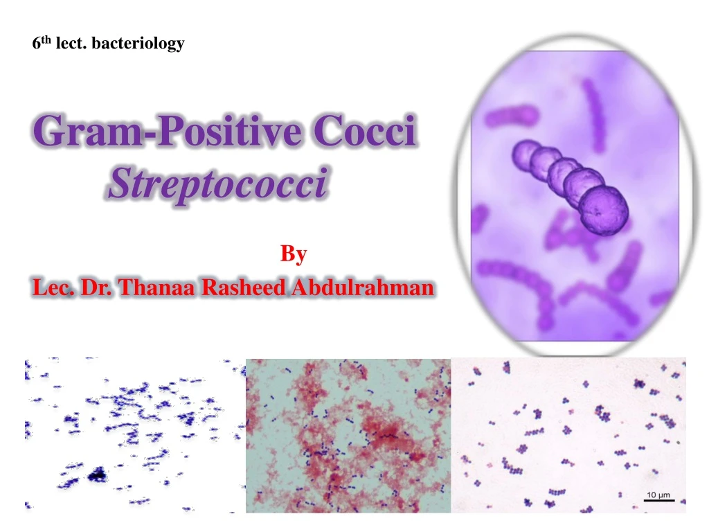
6th lect. bacteriology Gram-Positive CocciStreptococci By Lec. Dr. ThanaaRasheedAbdulrahman
Streptococci of medical importance are listed in Table 1 1All streptococci are catalase-negative. 2S. bovis is a nonenterococcal group D organism. 3Both Ent. faecalis and Str. bovis grow on bile-esculin agar, whereas other streptococci do not. They hydrolyze the esculin, and this results in a characteristic black discoloration of the agar. 4NA, not applicable. 5Viridans group streptococci include several species, such as Str. sanguis, Str. mutans, Str. mitis, Str. gordoni, Str. salivarius, Str. anginosus, Str. milleri, and Str. intermedius.
Characteristics: They are non-motile, non-sporulating, gram- positive facultative anaerobes Spherical or oval cells characteristically forming pairs or chains during growth. Grow well on ordinary solid media enriched with blood, serum or glucose. Most streptococci grow in solid media as discoid colonies Capsular streptococcal strains give rise to mucoid colonies They are aerobic bacteria in which growth is enhanced with 10% carbondioxide. They are catalase-negative. They are widely distributed in nature and are found in upper respiratory tract, gastrointestinal tract and genitourinary tract as normal microbial flora. They are heterogeneous group of bacteria, and no one system suffices to classify them.
The currently used classification is based on : (1) Colony morphology and hemolytic reactions on blood agar; (2) Serologic specificity of the cell wall group-specific substance and other cell wall or capsular antigens; (3) Biochemical reactions and resistance to physical and chemical factors. (4) Ecologic features. (5) Molecular genetics have also been used to study the streptococci.
One of the most important characteristics for identification of streptococci is the type of hemolysis. • α-Hemolytic streptococci form a green zone around their colonies as a result of incomplete lysis of red blood cells in the agar. • β-Hemolytic streptococci form a clear zone around their colonies because complete lysis of the red cells occurs. β -Hemolysis is due to the production of enzymes (hemolysins) called streptolysin O and streptolysin S. • Some streptococci are nonhemolytic (γ-hemolysis).
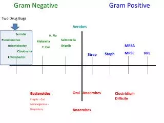
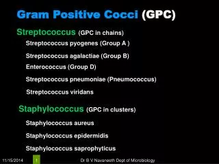
Classification of streptococciLancefield grouping of streptococci: • Streptococci produce group specific carbohydrates(Ccarbohydrates) identified using group specific antiserum. • It is designated A-H and K-V. • The clinically important streptococci are grouped under A,B,C,D, F and G. • The main species and groups of medical importance • S. pyogenes……... Lancefield group A • S. agalactiae…….. Lancefield group B • Enterococci………Lancefield group D • NB: Viridans streptococci and anaerobic streptococci are not grouped under Lancefield Classification.
Virulence factorsAntigenic structure: • Group-specific cell wall antigen Streptococcal cell wall obtained carbohydrate is the basis for serologic grouping of streptococci (Lancefield groups A-H, K-U).Its determine the group of β-hemolytic streptococci. It is specificity is determined by an amino sugar. 2. M protein is the most important virulence factor • They are found in hair-like projections of the streptococcal surface, protrudes from the outer surface ,determine virulence.It interferes with ingestion by phagocytes, i.e., it is antiphagocytic. Antibody to M protein provides type-specific immunity. • It’s a major virulent factor for group A streptococci. • There are two major structural classes of M protein(class I &class II) and more than eighty serotypes of M protein of group A streptococci • The class I M protein may be a virulence determinant for rheumatic fever • Conserved antigenic domains on the class I M protein induce antibodies that cross react with cardiac muscle tissue. • Strains of Str. pyogenes that produce other M protein types are nephritogenic, i.e., cause primarily acute glomerulonephritis.
Antigenic structure 3. Polysaccharide capsulethat plays a role in retarding phagocytosis 4. T substance: Acid and heat labile unlike M protein, and has no relation to virulence of streptococci. 5. R protein. Transmission Most streptococci are part of the normal flora of the human throat, skin, and intestines but produce disease when they gain access to tissues or blood. Viridans streptococci and Str. pneumoniae are found chiefly in the oropharynx; Str. pyogenes is found on the skin and in the oropharynx in small numbers; Str. agalactiae occurs in the vagina and colon; and both the enterococci and anaerobic streptococci are located in the colon.
(Group A β-hemolytic streptococci) Streptococcus pyogenes • The most pathogenic member of the genus • It is present as a commensal in the nasopharynx in a variable proportion of healthy individuals. • Cause pharyngitis and a very common cause of skin infections. • They adhere to pharyngeal epithelium via pili covered with lipoteichoic acid and M protein. • Many strains have a hyaluronic acid capsule that is antiphagocytic. • The growth of Str. pyogeneson agar plates in the laboratory is inhibited by the antibiotic bacitracin, an important diagnostic criterion. • It produces different types of enzymes and exotoxins.
Pathogenesis • Group A streptococci (Str. pyogenes) cause disease by three mechanisms: • Pyogenic inflammation. • Exotoxin production. • Immunologic, • Group A streptococci produce three important inflammation-related enzymes: • Hyaluronidase degrades hyaluronic acid, which is the ground substance of subcutaneous tissue. Hyaluronidase is known as spreading factor because it facilitates the rapid spread of Str. pyogenes in skin infections (cellulitis). • Streptokinase (fibrinolysin) activates plasminogen to form plasmin, which dissolves fibrin in clots, thrombi, and emboli. • DNase (streptodornase) degrades DNA in exudates or necrotic tissue. Antibody to DNase B develops during pyoderma; this can be used for diagnostic purposes.
Group A streptococci produce five important toxins and hemolysins: • Erythrogenictoxin (Pyrogenic toxin)causes the rash of scarlet fever. Its mechanism of action is similar to that of the toxic shock syndrome toxin (TSST) of Sta. aureus,i.e., it acts as a superantigen. It is produced by certain strains of Str. pyogeneslysogenized by a bacteriophage carrying the gene for the toxin. • The injection of a skin test dose of erythrogenic toxin (Dick test) gives a positive result in persons lacking antitoxin (i.e., susceptible persons). • StreptolysinOis a hemolysin that is inactivated by oxidation(oxygen-labile). It causes β-hemolysis only when colonies grow under the surface of a blood agar plate. It is antigenic, and antibody to it (ASO) develops after group A streptococcal infections. The titer of ASO antibody can be important in the diagnosis of rheumatic fever.
Streptolysin S is a hemolysinthat is not inactivated by oxygen (oxygen-stable). It is not antigenic but is responsible forβ -hemolysis when colonies grow on the surface of a blood agar plate. • Pyogenicexotoxin Ais the toxin responsible for most cases of streptococcal toxic shock syndrome. It has the same mode of action as does staphylococcal TSST, i.e., it is a superantigen that causes the release of large amounts of cytokines from helper T cells and macrophages. • ExotoxinB is a protease that rapidly destroys tissue and is produced in large amounts by the strains of Str. pyogenes, the so-called "flesh-eating" streptococci that cause necrotizing fasciitis.
Clinical features • Acute streptococccalsorethroatPharyngitischaracterized by inflammation, exudate, fever, leukocytosis, and tender cervical lymph nodes. If untreated, spontaneous recovery occurs in 10 days, but rheumatic fever may occur. • Untreated pharyngitis may extend to the middle ear (otitis media), the sinuses (sinusitis), the mastoids (mastoiditis), or the meninges (meningitis). • If the infecting streptococci produce erythrogenic toxin and the host lacks antitoxin, scarlet fever may result. A "strawberry" tongue is a characteristic lesion. • Str. pyogenes also causes streptococcal toxic shock syndrome. Streptococcal TSS typically has a recognizable site of pyogenic inflammation and blood cultures are often positive, whereas staphylococcal TSS typically has neither a site of pyogenic inflammation nor positive blood cultures.
Clinical features • Group A streptococci cause skin and soft tissue infections, such as cellulitis, erysipelas, necrotizing fasciitis (streptococcal gangrene)which is the extensive and rapidly spreading necrosis of skin and subcutaneous tissue • Impetigo, a form of pyoderma, is a superficial skin infection characterized by "honey-colored" crusted lesions. • Lymphangitis can occur, especially on the forearm associated with an infection on the hand. • Group A streptococci also cause endometritis (puerperal fever), septicemia originating in the infected uterus,a serious infection of pregnant women, and sepsis.
Post-streptococcal diseases: Immunological diseases • Acute rheumatic fever • Immunological damage to the heart valves and muscle following Streptococcal upper respiratory tract infection.The inflammation is caused by an immunologic (antibody) response to streptococcal M proteins that cross-react with human tissues. • It clinically presents with fever, malaise, migratory nonsppurativepolyarthritis, carditis, erythemamarginatum and subcutaneous nodules. • Note that these diseases appear several weeks approximately 2 weeks after the actual infection because that's the length of time it takes to produce sufficient antibodies. • ASO (1/200) titers and the erythrocyte sedimentation rate are elevated. • Most cases of pharyngitis caused by group A streptococci occur in children aged 5 to 15 years and hence rheumatic fever occurs in that age group.
Acute glomerulonephritis • Immunological damage to the kidney following infection of skin with certain M protein (e.g., M protein type 49 ) types of Str. pyogenes. • Typically occurs 2 to 3 weeks after skin infection in children. • The most striking clinical features are hypertension, edema of the face (especially periorbital edema) and ankles, and "smoky" urine (due to red cells in the urine). • Most patients recover completely. Reinfectionwith streptococci rarely leads to recurrence of glomerulonephritis. • The disease is initiated by antigen–antibody complexes on the glomerular basement membrane, and soluble antigens from streptococcal membranes may be the inciting antigen. • It can be prevented by early eradication of nephritogenic streptococci from skin colonization sites but not by administration of penicillin after the onset of symptoms.
Group B streptococci Str. agalactiae • Colonize the genital tract of some women. • They are usually bacitracin-resistant. They hydrolyze (break down) hippurate, an important diagnostic criterion. Clinical features • Neonatal sepsis, pneumonia, and meningitis The main predisposing factor is prolonged (longer than 18 hours) rupture of the membranes in women who are colonized with the organism and lack antibody to group B streptococci and who consequently are born without transplacentallyacquired IgGs. • This organism also causes pneumonia, endocarditis, arthritis, and osteomyelitis in adults. • Postpartum endometritis also occurs. • Diabetes is the main predisposing factor for adult group B streptococcal infections
Group D streptococci • Include Enterococci • and Nonenterococci (e.g., Str. bovis). • Enterococci(e.g., Ent. faecalis and Enterococcusfaecium) • Are members of the normal flora of the colon and are noted for their ability to cause urinary, biliary, and cardiovascular infections. • They are very hardy organisms; they can grow in hypertonic (6.5%) saline or in bile and are not killed by penicillin G. As a result, a synergistic combination of penicillin and an aminoglycoside (such as gentamicin) is required to kill enterococci.
Vancomycin can also be used, but vancomycin-resistant enterococci (VRE) have emerged and become an important cause of life-threatening nosocomial infections. • More strains of Ent. faecium are vancomycin-resistant than are strains of Ent. faecalis. Clinical features • Frequent cause of nosocomial infection(urinary tract infections) Indwelling urinary catheters and urinary tract instrumentation are important predisposing factors. • Sub acute bacterial endocarditis • They also cause intra-abdominal abscess and pelvic infections, typically in combination with anaerobes.
Nonenterococcal group D streptococci • Str. bovis, can cause similar infections but are much less hardy organisms, e.g., they are inhibited by 6.5% NaCl and killed by penicillin G. Causes endocarditis, especially in patients with carcinoma of the colon. • The hemolytic reaction of group D streptococci is variable: most are α-hemolytic, but some are β-hemolytic and others are non hemolytic. • Groups C, E, F, G, H, and K–U streptococci infrequently cause human disease.
Non–β-Hemolytic Streptococci • Some produce no hemolysis; others produce α-hemolysis. • The principal α-hemolytic organisms are Str. pneumoniaeand the viridans group of streptococci. • The viridans streptococci are not bile-soluble and not inhibited by optochin—in contrast to Str. pneumoniae, which is bile-soluble and inhibited by optochin.
Viridans streptococci Include:Streptococcus mitisStreptococcus mutansStreptococcus salivariusStreptococcus sanguis • Viridansstreptococci are part of the normal flora of the human pharynx and intermittently reach the bloodstream typically after dental surgery to cause infective endocarditis. • Str. mutans synthesizes polysaccharides (dextrans) that are found in dental plaque and lead to dental caries. • Streptococcus intermedius and Streptococcus anginosus (also known as the Str. anginosus-milleri group) are usually α-hemolytic or nonhemolytic, but some isolates are β-hemolytic. They are found primarily in the mouth and colon. Cause brain abscesses and abdominal abscesses.
Peptostreptococci • These grow under anaerobic or microaerophilic conditions and produce variable hemolysis. • Peptostreptococci are members of the normal flora of the gut, mouth, and the female genital tract and participate in mixed anaerobic infections. The term "mixed anaerobic infections" refers to the fact that these infections are caused by multiple bacteria, some of which are anaerobes and others are facultatives. • For example, peptostreptococci and viridansstreptococci, both members of the oral flora, are often found in brain abscesses following dental surgery. • Peptostreptococcusmagnus and Peptostreptococcusanaerobius are the species frequently isolated from clinical specimens.
Laboratory Diagnosis • Specimen: • S. pyogenes- Throat swab, pus, blood • S. agalactiae- High vaginal swab of women; blood and cerebrospinal fluid of new born • Enterococci- Blood, pus • Viridans streptococci- Blood • Smear: Non-motile gram-positive cocci in chains • Culture, biochemical tests and sensitivity testing: • Grow in aerobic and anaerobic environment at a temperature of 35-37 0c. • Grow in ordinary media with shiny or dry colonies with grey-white or colorless appearance. • S. pyogenes- • Shows small, translucent β-hemolytic colonies in 18 to 48 hours (clear zone of hemolysis in blood agar plate). • Does not grow in macConkey agar plate. • Bacitracin sensitive. • PyrrolidonylArylamidase (PYR) Test: Positive
Laboratory Diagnosis • S. agalactiae- • Shows clear zone of hemolysis in blood agar plate. • May show double zone of hemolysiswhenincubated anaerobically • Active in hippurate hydrolysis • CAMP test is used to identify the bacteria. Depend on the production of a protein that causes enhanced hemolysis on sheep blood agar when combined with β-hemolysin of Sta. aureus (CAMP test). • Viridans streptococci- • Show greenish discoloration of blood agar plate. • Optochinresistant. • Not soluble in bile salts. • Do not ferment inulin.
Laboratory Diagnosis • Enterococci- • Non-hemolytic or α hemolytic changes in blood agar plate. • Grow in the presence of 6.5% NaCl. • Grows in macconkey agar • Identified by litmus milk reduction test. • PYR-Positive • Bile esculin-positive ( hydrolyze esculin in the presence of bile, i.e., they produce a black pigment on bile-esculin agar.
Laboratory Diagnosis • The rapid test provide a diagnosis in approximately 10 minutes were developed. This test detects bacterial antigens in a throat swab specimen. • In the test, specific antigens from the group A streptococci are extracted from the throat swab with certain enzymes and are reacted with antibody to these antigens bound to latex particles. • Agglutination of the colored latex particles occurs if group A streptococci are present in the throat swab. • The specificity of these tests is high but the sensitivity is low, i.e., false-negative results can occur. • If the test result is negative but the clinical suspicion of streptococcal pharyngitis is high, a culture should be done. • A rapid test is also available for the detection of group B streptococci in vaginal and rectal samples. It detects the DNA of the organism and results can be obtained in approximately 1 hour
Laboratory Diagnosis Serologic: ASO titers are high soon after group A streptococcal infections. In patients suspected of having rheumatic fever, an elevated ASO titer is typically used as evidence of previous infection because throat culture results are often negative at the time . Titers of anti-DNase B are high in group A streptococcal skin infections and serve as an indicator of previous streptococcal infection in patients suspected of having AGN.
Treatment: • Group A streptococcal infections can be treated with either penicillin G or amoxicillin, but neither rheumatic fever nor AGN patients benefit from penicillin treatment after the onset of the two diseases. • In mild group A streptococcal infections, oral penicillin V can be used. • In penicillin-allergic patients, erythromycin or one of its long-acting derivatives, e.g., azithromycin, can be used. However, erythromycin-resistant strains of Str. pyogenes have emerged . • Clindamycincan also be used in penicillin-allergic patients. • Enterococci…………. Penicillin or vancomycin+ Gentamicin • Enterococciresistant to multiple drugs, e.g., penicillins, aminoglycosides, and vancomycin and VRE, two drugs are being used to treat infections: linezolid (Zyvox) and daptomycin (Cubicin). • Endocarditiscaused by most viridans streptococci is curable by prolonged penicillin treatment. • Group B streptococcal infections ,Str. bovis and Peptostreptococcitreated with penicillin G.
PREVENTION • Rheumatic fever can be prevented by penicillin usually with benzathine penicillin once each month for several years. There is no evidence that patients who have had AGN require similar penicillin prophylaxis. • The incidence of neonatal sepsis caused by group B streptococci can be reduced by a two-pronged approach: (1) All pregnant women at 35 to 37 weeks' gestation should be screened by doing vaginal and rectal cultures. If cultures are positive, then penicillin G (or ampicillin) should be administered intravenously at the time of delivery. (2) If the patient has not had cultures done, then penicillin G (or ampicillin) should be administered intravenously at the time of delivery to women who experience prolonged (longer than 18 hours) rupture of membranes, whose labor begins before 37 weeks' gestation, or who have a fever at the time of labor.
Streptococcus pneumoniae • Fastidious, lancet-shaped gram positive diplococci. • Possess a capsule of polysaccharidethat permits typing with specific antisera. • Found as a normal flora in the upper respiratory tract. • On blood agar they produce α-hemolysis. • In contrast to viridans streptococci, they are lysed by bile or deoxycholate and their growth is inhibited by optochin.
Antigenic structure • Capsular polysaccharide: Pathogenicitydeterminant with anti-phagocyticproperty. There are more than 85 serotypes of the bacteria based on capsular typing.Identified by capsule swelling test (quellungreaction). • C substance: Cell wall associated antigen • Protein M antigen • IgA1 protease: Enzyme which cleaves IgA1 Clinical features: • Lobar Pneumonia, Otitis media, Sinusitis • Bacteremia…Meningitis, Endocarditis, Septic arthritis. • Pneumococci are the most common cause of community acquired pneumonia, meningitis, sepsis in splenectomized individuals. • They are a common cause of conjunctivitis, especially in children.
Laboratory Diagnosis: • Specimen:Sputum, blood, cerebrospinal fluid, ear discharge andsinus drainage. • Smears: Lancet-shaped gram positive diplococci • Culture: Grow best in chocolate agar media in CO2 enriched atmosphere. • Shows α-hemolytic, draughts man colony. • Appearance: Sunken centre colony due to spontaneous autolysis of older bacteria. • Young colonies resemble dew-drops due to capsule. • Bile soluble, ferment inulin. • Optochinsensitive. Serology: Quellungreaction Good for rapid identification ofS.pneumoniaein fresh specimen. • Procedure: Mix specific serotype ofS.pneumoniaewith specific anti-polysaccharide serum of the same serotype or with polyvalent anti-serum on a slide. Look for the appearance of capsule swelling under the 100X objective microscope
Treatment .Amoxicillin ,Chloramphenicol, Third generation Cephalosporins Prevention and control: • Pneumococcal conjugate vaccine to a carrier protein (diphtheria toxoid) : Immunization of individuals with type specific polysaccharide vaccine is given to children younger than 5 years and elderly (i.e., persons older than 65 years), immunocompromised (especially splenectomized) or debilitated persons. A booster dose is recommended . • Biochemical reaction to diagnose streptococci • Bile solubility test • Litmus milk reduction test • CAMP test • Bacitracin test • Optochintest