Download
1 / 37
390 likes | 971 Views
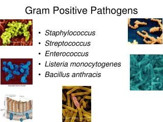
Important human pathogens of Gram-positive and Gram-negative cocci. SBM 2044 Medical Microbiology. Staphylococci. Staphylococci. Normal flora of the skin, nose, throat, and mucous membranes; cause suppuration, abscess, pyogenic infections, fatal septicaemia.

E N D
Important human pathogensof Gram-positive and Gram-negative cocci SBM 2044 Medical Microbiology
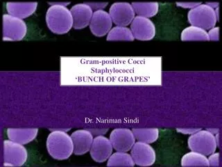
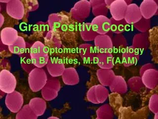
Staphylococci • Normal flora of the skin, nose, throat, and mucous membranes; cause suppuration, abscess, pyogenic infections, fatal septicaemia. • Haemolyse blood, coagulate plasma and produce a variety of extracellular enzymes and toxins • At least 30 species: S aureus, S epidermidis, S saprophyticus-UTI in young women • Staphylococci are non-motile; aerobic or microaerophilic and relatively resistant to drying and heat • S aureus is catalase +, coagulase +; form grey to golden yellow colonies, ferment mannitol.
Staphylococci • Staphylococci are resistant to drugs: • 1. β-lactamase production makes them resistant to penicillins (inc ampicillin) • 2. Independent β-lactamase production in resistant to nafcillin. mecA gene in chromosome. • 3. Resistant to vancomycin. Due to increased cell wall synthesis or alterations of cell wall, or the resistant vanA. • 4.Plasmid mediated resistance to tetracyclines, aminoglycosides.
Staphylococci - Antigenic Structure • Antigenic peptidoglycans (polysaccharides) stimulate production of interleukin-1 and opsonic antibodies; chemoattractant; endotoxin-like and activate complement. • Teichoic acids elicit anti-teichoic acid antibodies in endocarditis patients. • Protein A binds to the Fc portion of IgG molecules, agglutinate bacteria. • Capsules inhibit phagocytosis • Coagulase is a clumping factor on cell wall surface, binds to fibrinogen and yield bacterial aggregation.
Enzymes and toxins • 1. catalase • 2. coagulase and clumping factor – clots oxalated or citrated plasma. Coagulase binds to prothrombin, polymerize fibrin. • Hyaluronidase – spreading factor; staphylokinase – fibrinolysis; lipase. • Toxic shock syndrome toxin – TSST-1 is the superantigen; binds to MHC class II.
Regulations of vir determinants • Protein expressions depend on the bacterial growth phase. • Accessory global regulon gene, agr has 2 operons: 1st encodes RNA III that induces up-regulation of the secreted proteins and down-regulation of surface proteins.
Pathology • Furuncle or other localized abscess • Accumulation in a hair follicle→tissue necrosis. Coagulase coagulates fibrin, resulting in fibrous tissue. Center of the lesion turns to liquefaction of the necrotic tissue→granulation tissue→healing. • Suppuration via lymphatics or veins • Spreading of toxins, eg. TSST-1.
Clinical findings • Localised staph infection appears as “pimple” hair follicle infection, or abscess. • Dissemination of S. aureus→endocarditis, meningitis, pulmonary infection. 2° infection: symptoms like organ dysfunction or intense focal suppuration • Food poisoning due to staph enterotoxin presented with short incubation (1-8 hr); violent nausea, vomiting, diarrohea, rapid convalescence, no fever. • TSS manifested by abrupt onset of high fever, vomiting, diarrhoea, scarlatiniform rash, cardiac/renal failures. Women with tampons, or men and children with injured wounds. Virtually never in bloodstream.
MRSA = multiple resistant S aureus, because the organisms rapidly develop resistance to many antimicrobial drugs, drugs cannot act in the central necrotic part of a suppurative lesion. • Emergence resistant to erythromycin group – not used singly for treatment of chronic infection.
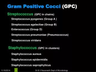
The Streptococci • Heterogenous group of bacteria, characterised by colony growth characteristics, haemolysis patterns on blood agar, antigenic composition of group-specific cell wall. • Spherical cocci in chains, Gram +. • Most group A, B and C produce hyaluronic acid capsules. • Cell wall contains proteins (M, T antigens), carbohydrates and peptidoglycans, . M-protein is hairl-like fimbriae.
Streptococci • Grow in blood or tissue fluids, 5% CO2 at 37°C. Group D enterococci grow well at 15°C and 45°C. • Classification is based on: group-specific cell wall Ag; M-protein; T-antigen and nucleoproteins. • -cell wall Ag by Lancefield grouping, by extraction of centrifuged culture with hot HCl, nitrous acid or formamide; by enzymatic lysis of strep cells (eg. Pepsin or trypsin).chemical structure of cell wall rhamnose acetylglucosamine. • - M-protein –virulent factor, able to resist phagocytosis by polymorphs. There are more than 100 serotypes of M proteins. M protein molecule rod-like coiled-coil structure. • -T antigen – not virulent, acid-labile and heat-labile.
Toxins and Enzymes • Streptokinase (fibrinolysin): human plasma→plasmin (active proteolytic enzyme that digests fibrin and other proteins). Rx of pulmonary emboli. • Hyaluronidase splits hyaluronic acid. Acts as a spreading factor • Haemolysins: streptolysin O (SLO)-inactivated in O2, anti-SLO level increases following infection (ASO serum titer of 160-200 units); and streptolysin S (SLS) – zones around colonies, elaborated in the presence of serum
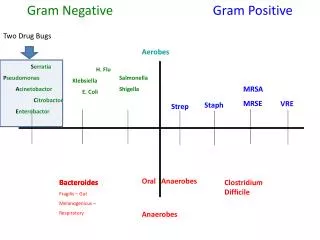
Streptococcus species • S pyogenes – Group A; β-haemolytic; human pathogen only; diseases such as local tonsillitis, necrotizing fasciitis and post-streptococcal glomerulonephritis. • S agalactiae- group B; β-haemolytic; NF of female genital tract, neonatal sepsis and meningitis. • Enterococcus faecalis – normal enteric flora; γ- or α-haemolytic. • S pneumoniae – α-haemolytic; growth is inhibited by optochin and colonies are bile-soluble. • Viridans streptococci - α-haemolytic typically; growth not inhibited by optochin and colonies are not bile-soluble; NF of the upper resp T; Viridans such as S mutans synthesise large polysaccharides (dextrans) contribute to dental caries.
Gram-negative bacilli Pseudomonads, vibrios, Helicobacter pylori, pasteurella and plague
Pseudomonads • Gram-negative bacilli; motile; aerobic; occur widely in soil, water, plants and animals. • P aeruginosa is an obligate aerobe, sometimes producing sweet or grape-like odour; forms smooth round colonies with fluorescent greenish pyoverdin in agar; bluish pigment pyocyanin. • In cystic fibrosis, P aeruginosa produces alginate, an exopolysaccharide that forms mucoid colonies and provide the matrix for biofilm. • Rx: combination of penicillin+aminoglycoside, not just a single-therapy because the bacteria can rapidly develop resistance with a single drug therapy.
P aeruginosa - toxins • Pili (fimbriae) promote attachment to epith. • Exopolysaccharide for mucoid colonies. • Lipopolysaccharide immunotypes; endotoxic. • Exotoxin A causes tissue necrosis by inhibiting protein synthesis (like diphtheria toxin).
Clinical findings • A nosocomial pathogen. • P aeruginosa infection gives rise to blue-green pus, meningitis (lumbar puncture) and UTI. • Organism is pathogenic only when introduced into areas devoid of normal defenses; when intravenous or urinary catheters are used; when neutropenia is present as in cancer chemo. • In most cases, symptoms and signs are nonspecific and are related to organ involved. • Ecthyma gangrenosum is haemorrhagic necrosis of the skin, where lesions are surrounded by erythema with no pus.
Burkholderia pseudomallei • Small, motile, aerobic G-, grows in 42°C. • Grows on standard media, colonies vary from mucoid and smooth to rough and wrinkled; from cream to orange in colour. • Causes melioidosis of humans, in Southeast Asia and N Australia. • -Incubation period is short (2-3 days); localised infection may result in acute septicaemic and may involve many organs. Commonly pulmonary infection (1° pneumonia)
Vibrio cholerae • Vibrios are comma-shaped aerobic rods, motile and possess a polar flagellum; oxidase +. • V cholerae produces enterotoxin that causes cholera (serogroups O1 and O139). • The culture presented as convex, smooth, opaque round colonies and granular; grow well at 37°C on thiosulfate-citrate-bile-sucrose agar with yellow colonies against the dark-green b/ground. Colonies are rapidly killed by acid.
Cholera • The connection between cholera and faecal bacteria in drinking water was discovered in London by John Snow in 1854. • Not an invasive infection. Organisms remain in intestinal tract. V cholerae attach to the microvilli of epith cells whereby they multiply and liberate cholera toxin. • Incubation period 1-4 days, presented with sudden onset of nausea, vomiting and profuse diarrhoea with abdominal cramps. Stools (“rice water”) contain mucus, epith cells and a lot of vibrios. • Rx: water and electrolyte replacement
Helicobacter pylori • A spiral-shaped G-; multiple flagella at one pole and highly motile; grow at 37°C pH 6-7 and killed in acidic; oxidase + and catalase +. • Cause gastritis, duodenal ulcer/peptic disease and gastric carcinoma. Urease producer. • Identification: H pylori grow in 3-6 days when incubated at 37°C in a microaerophilic environment. Colonies are translucent; 1-2mm in diameter.
H pylori - pathogenesis • H pylori ingested through food, retain in gastric mucus (physiological pH). • Produces: protease – makes mucous impermeable to acid; urease which yields ammonia production and buffers the acidic pH. • Presence of H pylori correlates with duodenal ulcer disease. • Lab tests: gastric biopsies, blood for serum Abs. • Immunity: Infected patients will develop IgM Abs. later, IgG and IgA. • Rx: Antibiotics with metronidazole, acid-suppressing agent for healing ulcers, proton pump inhibitors to inhibit organism (as well as urease inhibitors).
Plague • Plague – infection of wild rodents, transmitted interspecies, occasionally from rodents to humans, by bites of fleas. Outbreak seen in India, Africa, North/South of America and S.E.Asia. • Black death – pandemic that killed 2/3 of Europeans (~75mill people) in late 1340s. • Caused by Yersinia pestis, G- rod; exhibits striking bipolar staining with Wright-Giemsa stain; non-motile. • Antigenic structure: all possess LPS, which gives endotoxin activity when released.
Pathogenesis 1 Polymorphs→ killed Monocytes→ survive By coagulase
Clinical findings • After 2-7 days of incubation, patients presented with high fever, painful lymphadenopathy, enlarged, tender nodes (“buboes”: hence bubonic plague) in groin and axillae. Sometimes with vomiting and diarrhoea. • Later disseminated intravascular coagulation leads to hypotension, altered mental status, renal & cardiac failures. • Diagnostic: important to recognise early, confirms with lab. • Specimens taken from blood, aspirates of enlarged lymph nodes and sputum. • Control: (1) streptomycin, alternatively tetracycline; (2) destruction of plague-infected rodents, (3) vaccine (formalin-killed) for travellers.
Pasteurella • Gram - ; Non-motile; coccobacilli, aerobes or facultative anaerobes. • Pasteurella multocida – worldwide in Resp T and GIT; most common organisms in human wounds infected by bites from cats and dogs. • History of animal bite followed within hours of acute onset of redness, swelling and pain. • Rx: antibiotics penicillin G.
The Neisseriae • Gram – cocci; usually in pairs, sometimes piliated. 2 major species: gonococci and meningococci – differentiated by the usual clinical presentations of the diseases they cause. • Cultures: after 48h on enriched media (Mueller-Hinton) form convex, glistering, elevated, mucoid colonies, 1-5mm; non-haemolytic, transparent colonies. • Produce oxidase. Test: paper soaked with tetramethyl-paraphenylenediamine hydrochloride → dark purple rapidly. • Rapidly killed by drying, sunlight, moist, heat and many disinfectants.
Neisseria gonorrhoeae • Gonorrhoea "flow of seed"; in ancient times it was thought that the pus discharge associated with the disease contained semen. • Antigenic switching mechanism: pilin ↔Opa ↔ lipooligosaccharide (LOS) Rapid and occurs 1 in every 103 organisms Different mechanisms applied - Multiple genes encode for pilin,removal part of DNA for Opa.
Neisseria gonorrhoeae • Clinical specimens are looked for Opa protein; colonies form transparent or opaque. • Pathogenesis: gonococci attack mucous membranes of GUT, eyes, rectum: • Acute suppuration→tissue invasion→ chronic inflammation→fibrosis; spread to other tissues. • Gonococcal bacteriaemia leads to skin lesions, haemorrhagic papules and pustules. • Newborn – gonococcal ophthalmia neonatum.
Neisseria gonorrhoeae • Rx: localised infection – serum-sensitive (killed by Abs and c’) bacteria; whereas bacteriaemia – serum-resistant. • Diagnostic lab tests: taken from pus & secretions from urethra, cervix, throat, synovial fluid and smears from endocervix and urethral are stained. • Disease: worldwide, sexually, multiple sexual partners, asymptomatic infection.
Meningococcus • Piliated cocci; meningococci pathogenic to human only. • Symptoms with upper Resp T, high fever, sudden intense headache, vomiting, stiff neck, coma within hours. Meningococcemia – thrombosis of many small blood vessels in multiple organs. • Specimens: blood, spinal fluid, nasopharyngeal swab; all are tested with Gram staining and oxidase test. Further test: Abs to meningococcal polysaccharides by latex agglutination or haemagglutination.
PustulesPustules are well circumscribed elevations of the superficial layers of the epidermis. Like papules, they may be follicular or interfollicular in distribution. The most common cause is bacterial infection, where the pustules are filled with neutrophils, bacteria, debris, and possibly a few loose keratinocytes (acantholytic cells). It must be kept in mind that pustules, due to the thinness of the epidermis are usually transient. What is often observed is a transition from erythematous macules to papules to relatively few pustules to small crusts and scale. • Papules PPapules are circumscribed, solid elevations of the skin, up to 1 cm in diameter. The elevation may be due to accumulation of cells, fluid, debris or metabolic deposits and may be follicular or interfollicular in orientation. Papules often begin as erythematous macules.