Download
1 / 23
230 likes | 265 Views
Explore the composition of blood, cellular components, and histology of blood vessels and lymphatic organs. Learn about red blood cells, white blood cells, platelets, plasma, and the functions of various blood cells. Dive into the structure and function of arteries, veins, and lymphoid organs such as tonsils and the thymus. This comprehensive anatomy and histology guide is perfect for students in the Faculty of Pharmacy. Enhance your knowledge and understanding of the circulatory and lymphatic systems with detailed information and visual aids.

E N D
Blood, vessels, lymphaticorgans Faculty of Pharmacy 2ndHistologypractice Department of Anatomy, Histology and Embryology 2019.
Composition of blood CELLULAR COMPONENTS Red blood cells (erythrocytes) -99% of the cells -oxygen and carbondioxide transport White blood cells (leukocytes) -defence of the organism Platelets (thrombocytes) - blood clotting PLASMA -55 % of the blood -water -electrolytes -proteins albumin fibrinogen globulins -transported molecules (mostly transported bound to proteins) nutrients, vitamins, trace elements metabolic products hormones fatty substances
Blood smear and May-Grünwald Giemsa staining to study the morphology of blood cells one drop of blood
Cells in the blood smear erythrocytes monocyte eosinophil neutrophil basophil granulocytes neutrophil granulocyte small medium lymphocyte
Red blood cells (erythrocytes) -no cell nucleus -disk-shaped, concave on both sides, (special cytoskeleton with spektrin molecules) Function: O2 and CO2 transport lifespan: 120 days, mostly broken down in the spleen and in the liver (iron: stored, reused,
Platelets (thrombocytes) -cytoplasm fragments which derives from megakaryocytes (giant cells in the bone marrow) -lifespan: 5-10 days 250-300 000/mm3 2-3 mm Function: blood clotting
Neutrophil granulocyte -size: 10-15 mm -segmented nucleus (3-5lobes) -small granules in the citoplasm 60-70% of white blood cells also called phagocytes because they phagocytose foreign material (first cells to reach the site of an inflammation) part of the nonspecific immune system Neutrophil Chase https://www.youtube.com/watch?v=iFOus8ehxUc
Eosinophil granulocyte -size:10-15 mm; -two-lobed nucleus -eozinophil granules containing histamin, crystals can be detected byEM -1-6% of white blood cells -are capable of phagocytosis (Ag-Ab complexes), in allergic reactions they bound to and inactivate excess histamine (from mast cells or basophils)
Basophil granulocyte - size: 10-15 mm • segmented nucleus (not seen because of the granules) • many, large, basophil cytoplasmic granules (heparin, histamine). 0-1% of white blood cells effector cells in allergy, immediate hypersensitivity
Monocytes -size: 15-20 mm(largest WBC!) -ovale or kidney-shaped nucleus, numerous lysosomes in the cytoplasm -precursors of tissue macrophages 2-6 % of white blood cells Function: coordination of cellular and humoral immune response
Lymphocytes -size: 8-10 mm -round-shaped nucleus, organellum rich citoplasm -20-40 % of whit blood cells Functions: cells of the specific (humoral and cellular) immunity Subtypes: B- and T-lymphocytes NK cells (against tumor cells) activated lymphocytes increase their cytoplasm and rER
Histology of circulatorysystem • Tunicaintima • Tunicamedia: • Tunicaadventitia: capillary artery arteriole
Tonsils In the mucosa, local immunological protection Waldeyer’s lymphatic ring: pharynx, palate, auditory tube, root of the tongue Epithelial covering depends on the location and lymphoreticular tissue, no sinuses
General structure of the tonsils http://udspace.udel.edu/handle/19716/1999 Follicles Interfollicular area Tonsillar crypts Lymphoid infiltration with macrophages Reticular epithelium Discontinous lamina basalis
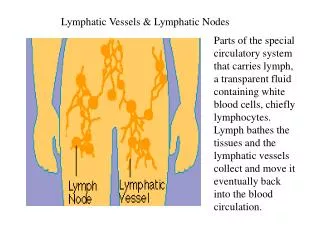
Lymph flow: aff. lymph vessels, subcapsular sinuses, cortical sinuses, intermediate or trabecular sinuses, medullary sinuses, terminal sinuses. During the passage the lymph is filtered and modified by the immune cells. Finally lymph is collected by the efferent lymphatics.
THYMUS HISTOLOGY CT capsule, trabeculae, incomplete lobules, cortex stains more darkly
37. Bloodsmear(MGG) eosinophil neutrophil RBC platelet monocyte lymphocyte
19. Artery and vein(HE) artery vein tunicaintima tunicamedia tunicaadventitia