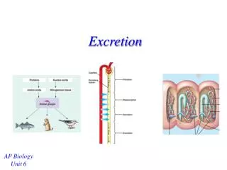
Excretion
Excretion. Animals must regulate the chemical composition of its body fluids by balancing the uptake and loss of water and fluids.

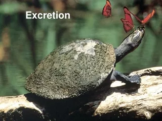
Excretion
E N D
Presentation Transcript
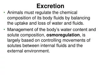
Excretion • Animals must regulate the chemical composition of its body fluids by balancing the uptake and loss of water and fluids. • Management of the body’s water content and solute composition, osmoregulation, is largely based on controlling movements of solutes between internal fluids and the external environment.
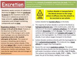
Animals must also remove metabolic wastes before they accumulate to harmful levels: • Water • Carbon dioxide • Salts • Bile pigments • Nitrogenous waste • Ammonia • Urea • Uric Acid • Creatine
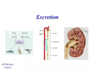
Metabolic Wastes • Because most metabolic wastes must be dissolved in water when they are removed from the body, the type and quantity of waste products may have a large impact on water balance. • In general, the kinds of nitrogenous wastes excreted depend on an animal’s evolutionary history and habitat - especially water availability. • The amount of nitrogenous waste produced is coupled to the energy budget and depends on how much and what kind of food an animals eats.
Animals that excrete nitrogenous wastes as ammonia need access to lots of water. • This is because ammonia is very soluble but can only be tolerated at very low concentrations. • Therefore, ammonia excretion is most common in aquatic species. • Many invertebrates release ammonia across the whole body surface. • In fish, most of the ammonia is lost as ammonium ions (NH4+) at the gill epithelium.
Ammonia excretion is much less suitable for land animals and even many marine fishes and turtles. • Because ammonia is so toxic, it can only be transported and excreted in large volumes of very dilute solutions. • Most terrestrial animals and many marine organisms (which tend to lose water to their environment by osmosis) do not have access to sufficient water. • Instead, mammals, most adult amphibians, and many marine fishes and turtles excrete mainly urea.
Urea is synthesized in the liver by combining ammonia with carbon dioxide and excreted by the kidneys. • The main advantage of urea is its low toxicity, about 100,000 times less than that of ammonia. • Urea can be transported and stored safely at high concentrations. • This reduces the amount of water needed for nitrogen excretion when releasing a concentrated solution of urea rather than a dilute solution of ammonia.
The main disadvantage of urea is that animals must expend energy to produce it from ammonia.
Land snails, insects, birds, and many reptiles excrete uric acid as the main nitrogenous waste. • Like urea, uric acid is relatively nontoxic. • But unlike either ammonia or urea, uric acid is largely insoluble in water and can be excreted as a semisolid paste with very small water loss. • While saving even more water than urea, it is even more energetically expensive to produce.
Human Excretory Organs • Lungs • Skin • Liver • Kidneys (Urinary system)
The Human Urinary System • Mammals have a pair of bean-shaped kidneys. • These are supplied with blood by a renal artery and a renal vein. • Urine exits each kidney through a duct called the ureter, and both ureters drain to a common urinary bladder. • During urination, urine is expelled from the urinary bladder through a tube called the urethra, which empties to the outside near the vagina in females or through the penis in males. • Sphincter muscles near the junction of the urethra and the bladder control urination.
The kidney has two distinct regions, an outer renal cortex and an inner renal medulla. • Both regions are packed with microscopic excretory tubules, nephrons, and their associated blood vessels. • Each human kidney packs about a million nephrons.
Nephron: Functional Unit of Kidney • Bowman’s capsule • Proximal convoluted tubule • Loop of Henle • Distal convoluted tubule • Collecting tubule
Circulatory System Interface with Urinary System • Renal artery • Renal arterioles • Afferent renal arteriole • Glomerulus Bowman’s Capsule • Efferent renal arteriole • Peritubular capillary network tubules • Renal venules • Renal vein
Nephron Peritubular capillary network
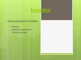
Stages of Urine Formation • Pressure Filtration : Glomerulus Bowman’s Capsule • Selective Reabsorption: Proximal convoluted tubule and Loop of Henle Peritubular capillary network • Tubular Secretion: Peritubular capillary network Distal convoluted tubule • Urine concentration: Collecting tubule
Pressure Filtration • Filtration occurs as blood pressure forces fluid from the blood in the glomerulus into the lumen of Bowman’s capsule. • The porous capillaries, along with specialized capsule cells called podocytes, are permeable to water and small solutes but not to blood cells or large molecules such as plasma proteins. • The filtrate in Bowman’s capsule contains salt, glucose, vitamins, nitrogenous wastes, and other small molecules.
Selective Reabsorption • From Bowman’s capsule, the filtrate passes through the proximal convoluted tubule and the loop of Henle, a hairpin turn with a descending limb and an ascending limb. • Selective reabsorption of molecules out of filtrate in nephron into blood in peritubular capillary network occurs in the proximal tubule
Valuable nutrients, including glucose, amino acids, and vitamins are actively or passively absorbed from filtrate into blood. • The epithelial cells actively transport Na+ out of the filtrate into the blood. • This transfer of positive charge is balanced by the passive transport of Cl- out of the filtrate into the blood. • As salt moves from the filtrate to the blood, water follows by osmosis.
The reabsorption of water continues as the filtrate moves into the descending limb of the loop of Henle. • The membrane of the descending limb is freely permeable to water but not very permeable to salt and other small solutes. • For water to move out of the tubule by osmosis, the interstitial tissue fluid bathing the tubule must be hypertonic to the filtrate. • Because the osmolarity of the interstitial tissue fluid does become progressively greater from the outer cortex to the inner medulla, the filtrate moving within the descending loop of Henle continues to loose water.
In contrast to the descending limb, the membrane of the ascending limb is permeable to salt, not water. • As filtrate ascends the thin segment of the ascending limb, NaCl diffuses out of the permeable tubule into the interstitial tissue fluid, increasing the osmolarity of the medulla. • The active transport of salt from the filtrate into the interstitial tissue fluid continues in the thick segment of the ascending limb, increasing the osmolarity of the medulla • This reinforces the water loss from the filtrate in the descending limb (counter-current effect)
Tubular Secretion and Urine Concentration • The distal tubule plays a key role in regulating the K+ and NaCl concentrations in body fluids by varying the amount of K+ that is secreted into the filtrate and the amount of NaCl reabsorbed from the filtrate. • The distal tubule also contributes to pH regulation by controlled secretion of H+ and the reabsorption of bicarbonate (HCO3-).
As the collecting duct traverses the gradient of osmolarity in the kidney from cortex to medulla, the filtrate becomes increasingly concentrated as it loses more and more water by osmosis to the hypertonic interstitial tissue fluid. • In the inner medulla, the collecting duct becomes permeable to urea, contributing to hypertonic interstitial tissue fluid and enabling the kidney to conserve water by excreting a hypertonic urine.
Hormones of the Kidney If blood pressure/ volume is too low: • Anti-diuretic Hormone (ADH) • Renin, Angiotensin II, Aldosterone If blood pressure/ volume is too high: • Natriuretic Peptide Hormones
One hormone important in regulating water balance is antidiuretic hormone (ADH). ADH is produced in hypothalamus of the brain and stored in and released from the pituitary gland, which lies just below the hypothalamus. Osmoreceptor cells in the hypothalamus monitor the osmolarity of the blood. Antidiuretic Hormone (ADH)
No ADH Present - Collecting tubule is NOT permeable to water and large volume of urine is produced Collecting tubule
ADH Present - Collecting tubule is permeable to water and a small volume of urine is produced Collecting tubule
Renin One of the functions of the kidney is to monitor blood pressure at the juxtaglomerular apparatus and take corrective action. If blood pressure should drop: • The juxtaglomerular apparatus of the kidney secretes the enzyme renin • Renin catalyzes the conversion of the plasma protein angiotensinogen to angiotensin I • Angiotensin converting enzyme (secreted by blood vessels) catalyzes the conversion of angiotensin I to angiotensin II
Angiotensin II • constricts the walls of arterioles increase blood pressure • stimulates the proximal convoluted tubules to reabsorb sodium ions water follows by osmosis increase blood pressure • stimulates the adrenal cortex to release the hormone aldosterone • aldosterone causes the kidneys to reclaim still more sodium ions water follows by osmosis increase blood pressure • increases the strength of the heartbeat • stimulates the pituitary glands to release ADH
Action of Renin, Angiotensin II, and Aldosterone Angiotensin I
Natriuretic Peptide Hormones • In response to a rise in blood pressure, the heart releases two peptides: • A-type Natriuretic Peptide (ANP) This hormone of 28 amino acids is released from stretched atria (hence the "A") • B-type Natriuretic Peptide (BNP) This hormone (29 amino acids) is released from the ventricles. (It was first discovered in brain tissue; hence the "B")
Both hormones lower blood pressure by • dilating arterioles • inhibiting the secretion of renin and aldosterone • inhibiting the reabsorption of sodium ions by the kidneys • The latter two effects reduce the reabsorption of water by the kidneys, so the volume of urine increases as does the amount of sodium excreted in it. The net effect of these actions is to reduce blood pressure by reducing the volume of blood in the circulatory system