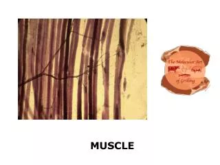
Muscle Overview
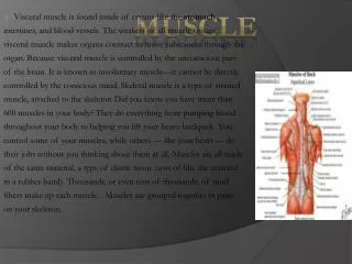
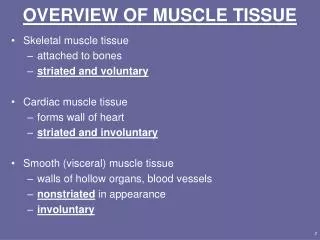
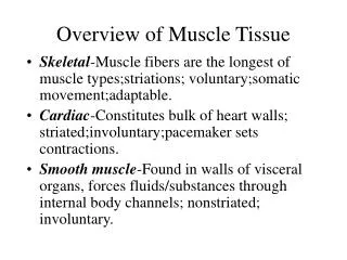
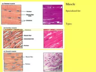
Muscle Overview. 3 different types of muscle tissue provide movement: Skeletal attached to the bones of the skeleton controlled consciously ( voluntary ) Cardiac heart controlled unconsciously ( involuntary ) Smooth

Muscle Overview
E N D
Presentation Transcript
Muscle Overview • 3 different types of muscle tissue provide movement: • Skeletal • attached to the bones of the skeleton • controlled consciously (voluntary) • Cardiac • heart • controlled unconsciously (involuntary) • Smooth • airways of the lungs, blood vessels, the digestive, urinary, and reproductive tracts • controlled unconsciously (involuntary)
Characteristics of Muscle Tissue • Excitability, or irritability • the ability to receive and respond to stimuli • Conductivity • the ability to create and conduct an action potential along the cell membrane • Contractility • the ability to shorten forcibly through the hydrolysis of ATP by contractile proteins • Extensibility • the ability to be stretched or extended • Elasticity • the ability to recoil after being stretched
Muscle Terminology • Prefixes • sarco-“flesh” • sarcolemma = muscle plasma membrane • sarcoplasm = cytoplasm of a muscle fiber (cell) • my- “muscle” • myocyte = muscle fiber • epimysium = the sheath of connective tissue that surrounds a skeletal muscle
Motor Unit: The Nerve-Muscle Functional Unit • A skeletal fiber will contract only after it is excited • A skeletal fiber is excited by the exocytosis of the neurotransmitter acetylcholine from a motor neuron at a synapse called the neuromuscular junction (NMJ) • generates a graded potential which can lead to an action potential in the fiber to trigger contraction • A single motor neuron is capable of stimulating multiple skeletal muscle fibers to contract simultaneously • one axon branches creating multiple axon termini • the anatomical relationship between a motor neuron and all skeletal fibers that it causes to contract is called a motor unit
Motor Unit: The Nerve-Muscle Functional Unit • The number of muscle fibers per motor unit can range: • few (small motor unit) • control fine movements (fingers, eyes) • several hundred (large motor unit) • control gross movements (arms, legs) • large weight-bearing muscles (back)
Muscle Twitch • The contraction followed by the relaxation of a muscle fiber to a single, brief threshold stimulus by a motor neuron is called a twitch • There are three phases of a muscle twitch • Latent (lag) period • time between the stimulation by a motor neuron and the beginning of contraction (few milliseconds) • Contractile period • contractile proteins within the fiber hydrolyze ATP causing the fiber to shorten resulting in an increase in tension (force) • Relaxation period • fiber lengthens causing tension to decrease
Contraction of Skeletal Muscle • The two types of muscle contractions are: • Isometric contraction = “samelength” • muscle contracts and produces tension, but does not shorten • trying to lift a car • Isotonic contraction = “sametension” • muscle contracts and produces tension • shortens as it contracts • lifting a pencil
Isometric Contractions • Tension increases to the muscle’s capacity, but the muscle neither shortens nor lengthens • Occurs if the load is greater than the tension the muscle is able to develop
Isotonic Contractions • In isotonic contractions, the muscle changes in length and moves the load
Types of Skeletal Muscle Fibers • There are 3 different types skeletal muscle fibers based on the duration of a twitch and the method of ATP production • slowoxidativefibers • fast oxidativefibers • fast glycolyticfibers • Skeletal muscles of your body contain a combination of all three fiber types, but their ratio determines the overall function of that muscle
Oxidative vs. Glycolytic fibers • Oxidative fibers contain greater amounts of mitochondria compared to glycolytic fibers • Oxidative fibers contain an oxygen-binding protein called myoglobin to maintain a high concentration of oxygen within the fiber for aerobic respiration • similar in structure to the blood protein hemoglobin • provides a red color to oxidative fibers • a lack of myoglobin in glycolytic fibers results in a white color
Characteristics of Skeletal Muscle Fiber Types • Slow oxidative fibers: • have a slow twitch (use ATP slowly) • fatigue resistant • muscle fibers used to maintain posture • Fast oxidative fibers: • have a fast twitch (use ATP quickly) • moderate resistance to fatigue • muscle fibers used for non-exertive movement (walking) • Fast glycolytic fibers: • have a fast twitch (use ATP quickly) • easily fatigued • muscle fibers used for powerful movements (jumping and sprinting)
Fatigue • Weakening of contracting muscle caused by: • the rate of ATP hydrolysis exceeds the rate of synthesis • lactic acid accumulation (↓ pH) inhibits muscle protein function • motor neurons run out of acetylcholine
Resistance to Fatigue • Fibers that use ATP slowly and have a high capacity to synthesize ATP are resistant to fatigue • Fibers that use ATP quickly and have a high capacity to synthesize ATP have moderate resistance to fatigue • Fibers that use ATP quickly and have a low capacity to synthesize ATP have no resistance to fatigue
Variety of Muscle Responses • Variations in the force of muscle contraction is required for proper control of skeletal movement • moving a pencil vs. a textbook with your hand uses the same muscles, but requires a different amount of force • Skeletal muscle contractions are varied by: • altering the number of muscle fibers that contract • determined by thenumber of motor units that are propagating action potentials to a muscle and which muscle fiber types are contracting to perform a particular task • altering the frequency of muscle stimulation • determined by the frequency of action potentials traveling down a motor neuron arriving at a fiber
Muscle Response: Motor Unit Recruitment • The first observable muscle contraction occurs following a threshold stimulus • activates one motor unit • As stimulus strength is increased more motor units are activated • recruitment • The maximum force that a muscle is capable of generating is reached when all motor units are activated • an increase in stimulus intensity results in no further increase in force generated
Motor Unit Recruitment • Slow oxidative fibers are first stimulated to contract • provide basal muscle tension (tone) • If additional muscle tension is required, fast oxidative fibers are stimulated to contract • Finally, the fast glycolytic fibers are stimulated to bring muscle tension to maximum
Muscle Response: Stimulation Frequency • Rapidly delivered stimuli result in the summation of muscle twitches creating an incomplete (unfused) tetanus (constant submaximal contractile force where each twitch is visibly distinct) • muscle tension does not return to baseline • If stimuli are given quickly enough, complete (fused) tetanus is observed where the contractile force reaches a maximum, but individual twitches blended together
ATP Sources During Muscle Contraction • Resting muscle fibers synthesizes and stores enough ATP (by cellular respiration) for 5 seconds of maximal sustained contraction. After that the muscle must make ATP in order to continue contraction • During resting periods, skeletal muscle uses ATP that it synthesizes to energize the amino acid derivative creatine into creatinephosphate which can be stored • during contraction creatine phosphate is converted back into creatine as ADP is converted to ATP • Glucose delivered to the muscle as well as stored glycogen (once hydrolyzed) is used by the muscle for additional ATP synthesis via glycolysis and oxidative phosphorylation
Monitoring of Muscle Length and Tension • Within skeletal muscle are 2 sensory receptors that monitor muscle length and tension • Muscle spindles are modified muscle fibers called intrafusal muscle fibers that are wrapped around by a neuron which sends information to the brain/spinal cord about the length of a muscle and the speed at which the length changes during contraction or stretching • extrafusal fibers are those that contract to produce tension and movement • Golgi tendon organs are neurons that are wrapped around the collagen fibers of a tendon near the attachment to muscle which sends information about the tension that a muscle produces during contraction
Muscle Spindles and Golgi Tendon Organs • Neurons associated with the spindle will generate additional or fewer APs which propagate to the brain/spinal cord when the length of the muscle (spindle) increases or decreases, respectively • Tension within a tendon (by either contraction or passive stretching) generates APs in the neuron which propagate to the brain/spinal cord
Muscle Spindles • A lengthened spindle generates more APs, a shortened spindle generates fewer APs • The brain/spinal cord interprets the change in the AP frequency from the spindle as a change in length
Myotatic Reflex • Reflex that causes the contraction of a muscle following an increase in that muscles length • APs from the lengthened spindle synapse with neurons in the spinal cord causing: • contraction of the extensors (pathway A and C) • relaxation of the opposing flexors (pathway B • sensory (pathway D) for perception by the brain
Golgi Tendon Organ • Tension within a tendon generates APs in the neuron which propagate to the brain/spinal • The greater the tension the higher the frequency of APs are generated so the brain/spinal cord can monitor the amount of stress in the tendon
Golgi Tendon Reflex • Protective reflex that prevents over contraction of a muscle resulting in damage to the muscle, tendon or bone • Contraction of the extensor muscle on the thigh stretches the Golgi tendon organ and generates APs causing: • inhibition of the motor neurons that innervate the extensor (A) • excitation in the opposing flexor’s motor neurons (B)
Microscopic Anatomy of a Skeletal Muscle Fiber • Each fiber is long (up to 30 cm) and cylindrical with multiple nuclei just beneath the sarcolemma • the sarcolemma contains both voltage-gated Na+ and K+ capable of generating an action potential • portions of the sarcolemma called transverse (t) -tubules fold inward toward the center of the fiber • propagate APs to the center of the muscle cell • Muscle fibers contain an elaborate, smooth sarcoplasmic (endoplasmic) reticulum(SR) • physically associated with the t-tubules • storage site of intracellular calcium (Ca+2) • An action potential in the t-tubules causes the release of from the SR into the sarcoplasm which increases the cytoplasmic level of Ca+2 • triggers the contraction of a muscle fiber
Contractile Proteins • Occupying most of the space within the cell, long filamentous contractile proteins are arranged in long bundles called myofibrils • composed of 2 types of contractile proteins (myofilaments) that overlap and slide past one another during contraction and relaxation • “thin” • “thick”
Structure of Thin Filaments • Thin filaments are composed of 3 proteins • F (fibrous) Actin is a helicalpolymer of G (globular) actin protein subunits • each subunit contains a binding site for the protein myosin of the thick filaments • Tropomyosin blocks the interaction between actin and myosin • prevents an unstimulated muscle from contracting • Troponin C is attached to tropomyosin • binds to Ca2+ in the sarcoplasm during contraction
Structure of Thick Filaments • Thick filaments are composed of many molecules of the protein myosin • Each myosin protein has a rodlike tail and two heads • Myosin heads: • hydrolyze a molecule of ATP • uses the chemical energy to contract • Temporarily bind to actin • pull on actin causing the shortening sarcomere
Striations of Skeletal Muscle • The overlapping arrangement of myofilaments creates a repeating pattern of striations (stripes) called sarcomeres when viewed longitudinally
Segments of a Sarcomere • Z disc • constitutes the end of a sarcomere • anchors the thin filaments • A band • the length of the thick filaments • I band • the length of thin filaments within a sarcomere that is not overlapping with the thick filaments • H (bare) zone • the length of thick filaments within in a sarcomere that is not overlapping with the thin filaments • During contraction, the thin and thick filaments slide past one another as the sarcomere shortens
Sliding Filament Model of Contraction • In the relaxed state, thin and thick filaments overlap only slightly • Upon stimulation, the thick filaments pull the thin filaments toward the center of the sarcomere • filaments overlap to a greater degree • shortening the sarcomere • As all of the sarcomeres in a muscle shortens, the entire muscle shortens
Skeletal Muscle Contraction • In order to contract, a skeletal muscle must be stimulated by a motor neuron • generates an action potential in the muscle fiber • causes an increase in the amount of cytoplasmic Ca2+ • causes the muscle fiber to contract • Linking the action potential to the contraction of a muscle fiber is called excitation-contraction coupling
Neuromuscular Junction • The axon termini have synaptic vesicles that contain the neurotransmitter acetylcholine(ACh) • ACh receptors (ligand-gated Na+ channels) are localized to a portion of the sarcolemma called the motor end plate
Excitation-Contraction Coupling • Binding of ACh to its receptors opens the channel and allows both Na+ and K+ to diffuse • diffusion of more Na+ than K+ causes the membrane potential to depolarize (endplate potential)