Download
1 / 35
350 likes | 489 Views
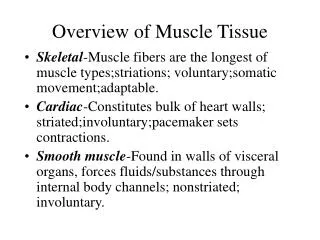
OVERVIEW OF MUSCLE TISSUE. Skeletal muscle tissue attached to bones striated and voluntary Cardiac muscle tissue forms wall of heart striated and involuntary Smooth (visceral) muscle tissue walls of hollow organs, blood vessels nonstriated in appearance involuntary.

E N D
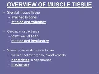
OVERVIEW OF MUSCLE TISSUE • Skeletal muscle tissue • attached to bones • striated and voluntary • Cardiac muscle tissue • forms wall of heart • striated and involuntary • Smooth (visceral) muscle tissue • walls of hollow organs, blood vessels • nonstriated in appearance • involuntary
Functions of Muscle Tissue • Produce body movements • Stabilize body positions • Storage/movement of substances within the body • bands of smooth muscle called sphincters • blood, lymph, urine, air, food and fluids, sperm • Heat production • involuntary contractions of skeletal muscle (shivering)
Properties of Muscle Tissue • Excitability = ability to generate electrical signals (a.p.’s) • in response to autorhythmic electrical signal in heart • in response to chemicals released from nerve cells (NTs) • Contractility • ability to contract forcefully when stimulated by act. pot. • Extensibility • ability to be stretched • can forcefully contract even when stretched • Elasticity = ability to return to original shape after being stretched
Skeletal Muscle: CT components • Superficial fascia= loose C.T. underlying the sub-Q layer of skin • Deep fascia = dense irregular C.T. around muscle • C.T. components of the muscle include • epimysium = surrounds whole muscle • perimysium = surrounds bundles (fascicles) of 10-100 muscle cells • endomysium = separates individual muscle cells • All these connective tissue layers extend beyond the muscle belly to form the tendon
Nerve and Blood Supply • Well-vascularized & highly innervated • Somatic motor neurons • axon extends from brain to group of fibers • axon branches @ muscle, each branch supplies diff. fiber • Each muscle cell supplied by at least one capillary • bring in oxygen/nutrients • remove wastes • Nerve fibers & capillaries are found in the endomysium between individual cells
Microscopic Anatomy of Skeletal Muscle • Muscle fibers = muscle cells • Sarcolemma = plasma membrane of muscle cell • T tubules = tiny in-folds of the sarcolemma • tunnel from surface of cell to center • open to outside of fiber filled with interstitial fluid • allows muscle action potential to propagate to all parts of muscle fiber simultaneously • Sarcoplasm = muscle cell cytoplasm • contains a large amount of glycogen for energy production • myoglobin binds oxygen for storage
Microscopic Anatomy of Skeletal Muscle • Sarcoplasmic reticulum • fluid-filled system of membranous sacs • similar to endoplasmic reticulum • encircles each myofibril • terminal cisternae store Ca+2 ions in relaxed muscle • release of Ca+2 ions triggers muscle contraction • intersection w/ T-tubule forms triad
Microscopic Anatomy of Skeletal Muscle • Myofibrils = contractile organelles of muscle fibers • extend entire length of muscle fiber • dark & light bands give striated appearance • sarcomere = functional unit of myofibril • contain myofilaments which do not extend entire length of cell • defined as the region btwn two Z discs • thin filaments • make up I band of sarcomere • actin protein • anchored together by Z disc in middle of I band • slide relative to myosin during muscle contraction
Microscopic Anatomy of Skeletal Muscle • Myofibrils, ctd. • thick filaments • myosin protein • “A” band of sarcomere dark-staining • “zone of overlap” = ends of A bands where thick/thin filaments overlap • H zone of sarcomere = region of only thick filaments • middle of A band where thin filaments do not reach • M line in center anchors myosin vertically
Filaments and the Sarcomere • Myofilaments do not extend entire length of muscle fiber arranged into sarcomeres • Sarcomere = functional unit of myofibril • Thick and thin filaments overlap each other in a pattern that creates striations (light I bands and dark A bands) • I band region contains only thin filaments • Separated by Z discs
Muscle Proteins • Contractile proteins generate force during muscle contraction • myosin = motor protein • push/pull cell structures to produce movement • actin = main component of thin filaments • myosin binding site on each molecule • Regulatory proteins • tropomyosin • covers myosin binding sites in relaxed muscle • prevents myosin binding in absence of Ca+2 ions • troponin holds tropomyosin in place • binds calcium • moves tropomyosin & exposes myosin binding sites • Structural proteins contribute to proper alignment, elasticity and extensibility
Sliding Filament Mechanism Of Contraction • Myosin cross bridges pullon thin filaments • Thin filaments slide toward M-line • Z Discs come toward each other • ***H & I bands narrow*** • Sarcomeres shorten at same time muscle fiber shortens muscle shortens • Notice: Thick/thin filaments do not change in length!!
How Does Contraction Begin? • Nerve impulse reaches axon terminal & synaptic vesicles release acetylcholine (ACh) • ACh diffuses across synaptic cleft & binds to receptors on the sarcolemma; Na+ channels open& Na+ rushes into cell • A muscle action potential spreads over sarcolemma and down into the transverse tubules • SR releases Ca+2 into the sarcoplasm • Ca+2 binds to troponin & causes troponin-tropomyosin complex to move & reveal myosin binding sites on actin contraction cycle begins
Contraction Cycle • Once Ca+2 is released from terminal cisternae & has bound troponin, myosin binding sites on actin are available for crossbridge formation • 4 steps to contraction cycle • ATP hydrolysis puts myosin in hi-energy position • attachment of myosin to actin to form crossbridges • Release of ADP & Pi causes conformational change in myosin which pulls thin filaments toward center of sarcomere this is the power stroke • detachment of myosin from actin when new ATP binds mysoin returns to hi-energy conformation (this is position of myosin in resting muscle) • Cycle repeats as long as ATP is available & [Ca+2] is high near the filaments
Steps in the Contraction Cycle Excitation - Contraction Coupling • Refer to figure 10.7 • Notice how myosin head attaches & pulls on thin filament with energy released from ATP • All the steps that occur from the muscle action potential reaching the T tubule to contraction of the muscle fiber • Refer to figure 10.8
Relaxation • Acetylcholinesterase (AChE) breaks down ACh @ synaptic cleft • Muscle action potentials cease • Ca+2-release channels close • Ca+2actively resequestered by SR/ Ca+2-ATPase pump • ↓ in sarcoplasmic Ca+2 returns troponin-tropomyosin to original conformation • myosin binding sites unavailable • Contraction stops but myosin heads remain in high-energy conformation, ready for contraction cycle to begin again ***Remember: ATP is required to detach crossbridges, so resting muscle is in energized state***
Length-Tension Relationship • Fiber length @ onset of contration influenes contraction strength • Maximal tension develops when fiber contracts @ + 30% of its optimal length • fibers shorter/longer than optimal length result in decreased force of contraction • too long: no overlap between myosin/actin no Xbridge formation • too short: too much overlap, thick filaments compressed by Z lines fewer interactions btwn myson/actin • Figure 10.9 illustrates this relationship
Structures of NMJ Region • NMJ = synapse btwn somatic motor neuron (SMN) & skeletal muscle fiber • synapse = area of communication btwn neuron & another cell (in this case, a muscle fiber) • synaptic cleft = gap separating neuron & muscle cell • neurotransmitter (NT) = chem released by neuron into cleft which activates the muscle fiber • axon terminal = end of motor neuron • synaptic end bulbs are swellings axon terminal • contain synaptic vesicles filled w/ ACh • motor end plate = region of sarcolemma opposite axon terminal • contains ACh receptors/cation channels
Generation of Muscle Action Potential • Arrival of nerve impulse at nerve terminal causes release of ACh from synaptic vesicles • ACh binds to receptors on motor end plate & opens gated ion channels Na+ rushes into muscle cell • Inside of muscle cell becomes more positive, triggering a muscle action potential that travels over the cell and down the T tubules • The release of Ca+2 from the SR is triggered and the muscle cell will shorten & generate force • Acetylcholinesterase breaks down the ACh attached to the receptors on the motor end plate so the muscle action potential will cease and the muscle cell will relax.
NMJ Antagonists • Botulinum toxin (C. botulinim) • blocks exocytosis of ACh muscle cannot contract • causes flaccid paralysis • death occurs from paralysis of the diaphragm • Curare (plant poison from poison arrows) • binds ACh receptors but does not activate them • causesflaccid paralysis • used to relax skeletal muscle during surgery because does not affect heart muscle • Black widow spider venom • causes massive ACh release • spastic paralysis results exaggerated reflexes, ↑ tone
Muscle MetabolismProduction of ATP in Muscle Fibers • Active muscle has high demand for ATP • required for contraction cycle • required for calcium uptake by SR • required for other metabolic reactions • Sarcoplasmic ATP only lasts for few seconds • 3 sources of ATP production within muscle • creatine phosphate • anaerobic cellular respiration • aerobic cellular respiration
Creatine Phosphate • Hi-energy compound found only in muscle • [Creatine phosphate] 3-6 times greater than [ATP] in muscle • Excess ATP within resting muscle used to form creatine phosphate • ATP + Cr ADP + Cr—P (at rest) • As ATP is used during exercise, [ADP] ↑ energy transferred from Cr—P to ADP • ADP + Cr—P ATP + Cr (in active muscle) • Sustains short, powerful bursts of maximal contraction • lasts about 15 sec • as in 100 meter dash
Anaerobic Cellular Respiration • Generates ATP w/o requiring O2 • When Cr—P used up, glucose broken down to form ATP • glycolysis yields 2 net ATP • pyruvate converted to lactic acid instead of entering aerobic metabolic cycles • Can provide ATP for 30 to 40 sec of maximal activity (200 meter race)
Aerobic Cellular Respiration • Series of reactions requiring O2 to generate ATP • If sufficient O2 is available, glc completely oxidized to yield 36 or 38 ATP • pyruvate enters mitochondria rather than being converted to lactate • 2 sources of oxygen • myoglobin • diffusion from blood • Provides ATP for prolonged activity if O2 & nutrients are available
Muscle Fatigue • Inability to contract after prolonged activity • Factors that contribute to fatigue: • central (psychological) fatigue = feeling of tiredness & desire to stop physical activity • neuromuscular fatigue = insufficient release of ACh from motor neurons • muscular fatigue: • depletion of creatine phosphate, glycogen • inadequate release of Ca+2 from SR • insufficient oxygen • lactate accumulation • Oxygen debt = amount of O2 required to replenish ATP/Cr—P used during exercise
The Motor Unit • Motor unit = one somatic motor neuron & all skeletal muscle cells (fibers) it stimulates • Muscle fibers of a unit normally scattered throughout belly of muscle (not all in one location) • All fibers within one unit contract in unison • Total strength of a contraction depends on # motor units activated & size of motor units • precise movements require large # of small motor units (few fibers/unit) • large/powerful movements involve large motor units (many fibers/unit)
Twitch Contraction • Brief contraction of all fibers in a motor unit in response to single action potential in its motor neuron • Latent period = delay btwn stimulus & onset of contraction • Muscle a.p. moving along sarcolemma & Ca+2 is released from SR • Contraction period = period where crossbridges form and peak muscle tension develops • Relaxation period = time during which Ca+2 is actively taken up by SR pump • Refractory period = period when muscle cannot contract even if it receives an additional stimulus
Frequency of Stimulation • Wave summation = repeated stimuli arriving @ different times, (but before muscle has completely relaxed from previous stimulus) produces an additive effect • results in stronger contraction • Incomplete (unfused) tetanus = sustained muscle contraction that permits partial relaxation between stimuli • stimulation rate ~20-30x/sec • Complete (fused) tetanus = sustained contraction that lackspartial relaxation between stimuli • stimulation rate ~ 80-100x/sec
Motor Unit Recruitment • Process in which # of active motor units is increased • Generally, motor units in a whole muscle fire asynchronously • some fibers are active & others are relaxed • Asynchronous contraction delays muscle fatigue allows for sustained contraction • Produces smooth muscular contraction • not series of jerky movements • Weak motor units are recruited first • then progressively larger units recruited as needed • size of unit recruited dependent upon amt of force needed
TYPES OF SKELETAL MUSCLE FIBERS • Slow oxidative fibers • Least powerful of three fiber types • Large amounts of myoglobin dark in color • Generate ATP primarily by aerobic cellular respiration • have many large mitochondria for this function • Highest resistance to fatigue of all fiber types adapted for endurance activities • Fast oxidative-glycolytic fibers • Lots of myoglobin, but less than slow oxidative fibers • Generate ATP via aerobic respiration • High glycogen content can also generate fair amt of ATP via anaerobic means • Fairly fatigue resistant
TYPES OF SKELETAL MUSCLE FIBERS • Fast glycolytic fibers • Generate quickest, most powerful contractions of all fiber types • Low myoglobin content appear white • Lots of glycogen primarily generate ATP via anaerobic means • Adapted for short, powerful movements weight lifting, throwing a ball, sprinting, etc. • Fatigue quickly • Relative ratios of fiber types are genetically determined • All muscles contain the three fiber types, but distribution offibers dependent upon particular function/need of the muscle
CARDIAC MUSCLE TISSUE • Found only in the heart wall • fibers arranged similarly to skeletal muscle fibers but are branched rather than parallel • fibers connect to adjacent fibers by intercalated discs • desmosomes prevent cell separation during contraction • gap junctions allow a.p. to spread quickly • contractions last longer than skeletal muscle twitch due to prolonged delivery of calcium ions from sarcoplasmic reticulum and the extracellular fluid. • Cardiac muscle fibers contract when stimulated by their own autorhythmic fibers. • This continuous, rhythmic activity is a major physiological difference between cardiac and skeletal muscle tissue.
SMOOTH MUSCLE • Nonstriated, involuntary & autorhythmic • Two types • Visceral (single unit) smooth muscle • found in walls of hollow viscera & small blood vessels; • fibers work as single unit, an a.p. stimulates all fibers to contract at once • Multiunit smooth muscle • found in large blood vessels, large airways, arrector pili muscles, eye muscles • each fiber has own neuron • fibers operate singly rather than as one unit
Microscopic Anatomy of Smooth Muscle • One centrally located nucleus • Thick and thin filaments are not organized into sarcomeres • Contains intermediate filaments attached to dense bodies • Small SR, but no T-tubules • Caveolae = structures containing XC Ca+2 for use by sm. musc. • Lengthwise shortening of fiber results from dense bodies pulling on filament • Twists into helix during contraction • Reverse motion during relaxation
Physiology of Smooth Muscle • Contraction starts slowly & lasts longer • no transverse tubules & very little SR • Ca+2 must flow in from outside • Can contract/stretch to greater extent than skeletal muscle • Regulatory protein = calmodulin • binds Ca in cytosol facilitates myosin-actin binding • Smooth muscle tone = state of c’td partial contraction • result of prolonged presence of calcium ions • Stress-relaxation response: smooth muscle can change in length without generating tension • retains contractile ability while stretched