Download

1 / 32
320 likes | 505 Views
Muscle Functions. Producing movement-Skeletal muscle is responsible for all somatic movements

E N D
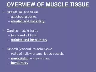
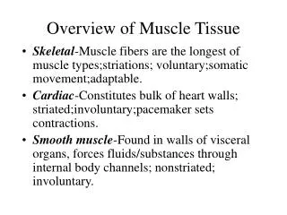
1. Overview of Muscle Tissue Skeletal-Muscle fibers are the longest of muscle types;striations; voluntary;somatic movement;adaptable.
Cardiac-Constitutes bulk of heart walls; striated;involuntary;pacemaker sets contractions.
Smooth muscle-Found in walls of visceral organs, forces fluids/substances through internal body channels; nonstriated; involuntary.
2. Muscle Functions Producing movement-Skeletal muscle is responsible for all somatic movements & manipulation;cardiac muscle courses blood through vessels;smooth muscle-peristaltic actions
Maintaining posture-Continuously defying gravity via constant adjustments
Stabilizing/strengthen joints
Generation of heat-Skeletal muscle contractions responsible for heat production.
3. Skeletal muscle functions Produce somatic movements
Maintain body posture/position
Reinforce soft tissue-anterior/posterior walls/pelvic floor
Guard entrances/exits-orifices of alimentary/urinary tracts
Regulation of body temperature-heat loss by muscle contractions
4. Gross Anatomy of Skeletal Muscles Epimysium-dense CT surrounds entire muscle;blends with deep fascia.
Perimysium and fascicles-fibrous CT surrounding bundles of fibers.
Endomysium-sheath of CT surrounding muscle fiber.
CT coverings contributes to muscle tissue elasticity
5. Nerve and Blood Supply Normal activity of skeletal muscle dependant on nerve/blood supply;each muscle is served by one nerve,artery & vein;nerves penetrate CT septa; each fiber supplied with nerve ending(myoneural junction).
Muscle requires large amount of energy;extensive blood supply delivers O2 & nutrients for ATP production.
6. Attachments Movable insertion moves towards immovable origin.
Direct attachment-epimysium fused to periosteum/perichondrium.
Indirect attachment-epimysium extends beyond muscles as sheet like aponeurosis; anchors muscle to bone, cartilage or fasciae of other muscles.
7. Microscopic Anatomy of Skeletal Muscle Muscle fiber is long, cylindrical, oval multinuclear myocyte; sarcolemma
Sarcoplasm contains glycosomes.
Myoglobin-red pigment
Myofibril-account for 80% of cellular volume.
8. Microscopic Anatomy of Skeletal Muscle (cont�d) Sarcoplasmic reticulum-membrane complex similar to SER; essential in contraction of myofibrils;t-tubule /terminal cisternae (triad)
Myofilaments-actin (thin filaments)/myosin (thick filaments); sarcomeres.
Sarcomere organization-M-line, Z-line, A-band (thick,thin), H band (thick), I-band (thin)
9. Tendons and Aponeuroses Tendon-fusion point of collagen fibers of endo-, peri-, and epimysium that attach muscle to bone, skin, or another muscle; resemble thick cords or cables
Aponeuroses-Formation of thick, flattened sheets.
10. Microscopic Anatomy of Skeletal Muscle (cont�d) Thin filaments-F-actin strand (G actin/tropomyosin, troponin.
Thick filaments-Myosin molecule (tail, head);elastic(titin)core
11. Naming Skeletal Muscles Location-temporalis, intercostals
Shape-deltoid, trapezius
Relative size-maximus, minimus, longus, brevis
Direction-rectus, oblique
Number of origins-biceps, triceps
Location of attachments-origin first/sternocleidomastoid
Action-flexor, extensor,adductor.
12. Arrangement of Skeletal Muscle Fibers Circular-orbicularis oris
Convergent-pectoralis major
Parallel-sartorius
Unipennate-extensor digitorum longus
Multipennate-deltoid
Fusiform-biceps
Bipennate-rectus femoris
13. The Axial Musculature The axial musculature is involved in moving the head and spinal column.
Categorized into five groups: (1) muscles of the head; (2) muscles of neck & vertebral column; (3)muscles of the thorax; (4) muscles of the abdominal wall;(5) muscles of the pelvic floor/perineum
14. Cephalic muscles Scalp
Epicranius-frontalis/occipitalis
Facial
Orbicularis oculi
Zygomaticus
Risorius
Orbicularis oris
Buccinator
Platysma
15. Cephalic Muscles (cont�d) Mastication
Temporalis
Medial/lateral pterygoids
Buccinator
Tongue movements
Genioglossus
16. Neck & Vertebral Column Neck
Sternocleidomastoid
Scalenes
Vertebral Column
Erector Spinae-spinalis,longissimus,
Iliocostalis
Semispinalis
Quadratus lumborum
17. Muscles of the Thorax External intercostals
Internal intercostals
Diaphragm
18. Muscles of the Abdominal Wall Rectus abdominis
External oblique
Internal oblique
Transversus abdominis
19. Muscles of the Pelvic Floor Levator ani- pubo-,iliococcygeus
Sphincter urethrae
Ischiocavernosus
Bulbospongiosus
20. Appendicular Skeletal Muscles Involved in stabilization of pectoral & pelvic limbs.
Accounts for 40% of skeletal muscle mass.
21. Pectoral Girdle Muscles/Upper Limbs 4 divisions
1. Pectoral girdle positioning
2. Brachium movement
3. Antebrachium movement
4. Hand/finger movement
22. Pectoral Girdle Positioning Anterior Thorax
Pectoralis minor
Serratus anterior
Posterior thorax
Trapezius
23. Brachium movement Pectoralis major
Latissimus dorsi
Deltoid
Subscapularis
Supra-,infraspinatus
Teres major, minor
Coracobrachialis
24. Antebrachium Movement Posterior aspect: extensors
Triceps
Anconeus
Anterior aspect: flexors
Biceps brachii
Brachialis
Brachioradialis
25. Hand/Finger Movement Anterior aspect
Pronator quadratus, teres
Flexor carpi radialis, ulnaris
Palmaris longus
Posterior aspect
Supinator
Extensor carpi radialis, ulnaris
Extensor digitorum
26. Intrinsic hand muscles Thenar (ball of thumb)
Hypothenar (ball of little finger)
Midpalm (lumbricals, interossei)
27. Pelvic Girdle Muscles and Lower Limbs Larger, stronger than pectoral limb muscles
3 groups: (1) Thigh movement; (2) Leg movement; (3) Foot & toe movement.
28. Thigh Movement Anterior compartment
Iliopsoas
Sartorius
Medial compartment
Adductor magnus, longus, brevis
Pectineus
Gracilis
29. Thigh Movement (cont�d) Posterior compartment
Gluteus maximus, medius, minimus
Piriformis
Obturator internus
Gemellus
Quadratus femoris
30. Leg Movement Anterior thigh compartment
Rectus femoris
Vastus lateralis, medialis, intermedius
Posterior thigh compartment
Biceps femoris
Semitendinosus
Semimembranosus
31. Foot & Toe Movement Anterior compartment
Tibialis anterior
Extensor digitorum longus
Extensor hallucis
Lateral compartment
Peroneus longus, brevis
32. Foot & Toe Movement (cont�d) Posterior compartment
Gastrocnemius
Soleus
Flexor digitorum longus
Tibialis posterior
Flexor hallucis longus