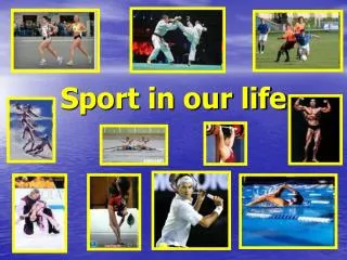
Sport & Soft tissues injuries
620 likes | 777 Views
Sport & Soft tissues injuries. Dr. Abdulaziz Alomar, MBBS, MSc , FRCSC Assistant professor Consultant O rthopaedic surgeon. Objectives . By the end of this teaching session the students should be able to

Sport & Soft tissues injuries
E N D
Presentation Transcript
Sport & Soft tissues injuries Dr. Abdulaziz Alomar, MBBS, MSc, FRCSC Assistant professor Consultant Orthopaedic surgeon
Objectives By the end of this teaching session the students should be able to • Specify the symptoms, signs and potential immediate complications of common sport and soft tissues injuries involving muscles, tendons, and ligaments for commonly injured joints; like shoulder, knee, and ankle. • Outline the assessment and appropriate investigation and to outline the immediate and long term management of patients with muscles, tendons, ligaments and meniscus • Demonstrate knowledge of indications for non-operative and operative treatment and to know the most common non-operative and operative measurements used for sport/soft tissue injuries.
Soft tissues injuries • Muscle • Tendon • Ligament • Meniscus • Knee • Shoulder • Ankle
R.I.C.E Compression Rest Elevation ICE
Muscle injury • The muscles most at risk are those in which the origin and the insertion cross two joints. • Frequently injured muscles act in an eccentric fashion (i.e., lengthening as they contract) • Frequently injured muscles have a relatively high percentage of type II (fast-twitch) fibers.
Muscle injuries • Muscle strain • Muscle Contusion • Muscle Laceration • Delayed-onset soreness
Muscle Strain • The most common muscle injury suffered in sports. • Immediate pain associated with diminished function. • Both complete and incomplete muscle tears can occur by passive stretch of an activated muscle. • Muscle tears also typically occur at or near to the myotendinous junction • Treatment • RICE • NSAID • physical therapy
Muscle Contusion • Caused by a nonpenetrating blunt injury (direct blow) to the muscle resulting in hematoma and inflammation. • Quadriceps and Brachialis muscles are common involved regions. • Clinical features: • Pain with active and passive motion +/_ swelling. • Decreased range of motion of joints spanned by the injured muscles. • Occasionally a permanent palpable mass. • Treatment: • Short period of immobilization • Followed by early mobilization and Physiotherapy • NSAID
Muscle injuries • Muscle Laceration • I&D followed by suture repair of the fascia, if possible. • Delayed-onset soreness • Structural muscle injury leads to progressive edema formation and resultant increased intramuscular pressure. • Is primarily associated with eccentric loading–type exercise. • Clinical features: muscular pain that occurs 1-3 days after vigorous exercise. • Treatment : • Will resolve in a few days • NSAID
Complications of muscle injures • Scar formation and muscle weakness. • Compartment syndrome • At the level of the muscle fibers, capillary bleeding and edema can lead to hematoma formation and can cause compartment syndrome in areas in which the volume is limited by the fascial envelope. • Pt with Bleeding disorders is at high risk • Myositis ossificans
Myositis ossificans • Bone formation within musclesecondary to blunt trauma. • CF: • Early: • Pain, swelling and decreased ROM • Erythema, warmth, induration, tenderness, • Late: painless swelling with decreased ROM • This sometimes mimics osteogenic sarcoma on radiographs and biopsy. • Increased ESR and serum alkaline phosphataseMyositis ossificans becomes apparent approximately 2 to 4 weeks post-injury.
Overuse Tendon injuries • Function—To transfer force from muscle to bone to produce joint motion. • Type of injuries • Overuse tendinopathies • Tendon rupture
Overuse tendinopathies • Osteotendinous junction is the most common site of overuse tendon injury. • Tendons are relatively hypovascular proximal to the tendon insertion.This hypovascularity may predispose the tendon to hypoxic tendon degeneration and has been implicated in the etiology of tendinopathies. • Tendinopathy not tendenitis
Most Common Diagnoses and Locations of Chronic Tendinopathies
Overuse tendinopathies treatment • Goal: reduce pain and return function. • Mainly is conservative Rx • Rest • Ice (Cryotherapy) • PT (stretching and eccentric strengthening) • Analgesics • Corticosteroids injection • Orthotics and braces • Other modalities: U/S, ESWT, iontophoresis and phonophoresis. • Surgical treatment: • Failed conservative treatment (at least 3-6 months) • Excision of abnormal tendon tissue and performance of longitudinal tenotomies to release areas of scarring and fibrosis.
Tendon rupture • Knee extensor mechanism • Quadriceps tendon • Patella tendon • Achilles tendon • Partial vs complete
Patella/Quadriceps tendon rupture • Predisposing factors: • Steroid, chronic disease, and tendinopathy • Age: Patella<40>Quads • Location: at the tendon attachment to the patella.
Patella/Quadriceps tendon rupture Physical examination: • Tenderness at the site of the injury,hematoma, and a palpable defect in the tendon. • Unable to extend the knee against resistance or to perform a straight-leg raise. • Xray • Patella-alta> P.T rupture • Patella-infera> Q.T rupture • Rx: usually surgical Patella-alta (high riding patella) seen with patella tendon rupture
Achilles tendon rupture • Most ruptures (75%) occur during sporting activities. • History: • The patient reports a “pop” or the sensation of being kicked in the heel during the injury. • weakness and difficulty walking. • Examination: • Increased resting dorsiflexion with the knees flexed, a palpable gap, weak plantar flexion, and an abnormal Thompson test (lack of plantar flexion when squeezing the calf). • Diagnosis is clinical, but MRI or ultrasound can confirm. • Rx: usually surgically
Knee • ACL • MCL • LCL • PCL • Menisci • Knee dislocation
ACL • Anatomy • Mechanism of injury • Diagnosis • Principles of management
Mechanism of Injury • Noncontact (70%) • Cutting or Pivoting • Contact = MCL • Sports-Related (80%) • “Pop” (70%) • Female: 2-4x > Male
Diagnosis • Symptoms: • Instability “giving way episodes” • Swelling (Hemarthrosis) is noted within a 1-2 days of the injury. • Pain if associated with meniscus tear
Diagnosis Physical examination • The patient need to be relaxed and comfortable. • Must be compared with those of the normal knee. • A moderate to severe effusion is usually present • ROM: in acute injury the range of motion may limited by: • Pain • Effusion • Hamstring spasm, • ACL stump impingement, • Meniscal pathology. • Special tests: • Lachman’s test • ADT • Pivot shift test : is pathognomonic for ACL injury (best in the chronic setting).
Diagnosis • Investigations: • X-ray • MRI • In the skeletally mature patient, the femoral insertion or midsubstance is usually the site of disruption. • In the skeletally immature patient, the tibial attachment may be avulsed with or without a piece of bone.`
Xray Segond fracture Tibial spine avulsion
MRI Normal ACL Torn ACL Bone bruise
INJURIES ASSOCIATED WITH ACL DISRUPTION • Injuries of the ACL rarely occur in isolation. The effects of other injuries, including: • Other ligament sprains (MCL) • Meniscal tears • Articular cartilage injuries • Bone bruises , • Complicate the treatment and eventual outcomes of ACL disruptions.
Treatment Nonsurgical treatment • Appropriate for asymptomatic patients with partial injuries to the ACL. • Patients who are older or less physically active may elect to modify their activities and proceed with nonsurgical treatment. If nonsurgical treatment fails or knee instability persists, surgery can be performed. • Nonsurgical treatment involves rehabilitation to strengthen hamstrings and quadriceps, as well as proprioceptive training. • Activity modification is also an important part of nonsurgical management, as patients who avoid cutting and pivoting sports are at lower risk for knee instability. • ACL sports braces are available as well. However, they have not been shown to prevent abnormal anterior tibial translation. Functional braces and simple knee sleeves improve proprioception, which may give patients a sense of improved knee function and stability.
Treatment Surgical • Athletes with ACL injuries rarely return to cutting and pivoting sports, such as basketball, football, soccer, squash, and handball, without first undergoing surgery. For individuals who wish to return to such sports, surgery is generally recommended to avoid instability and secondary meniscal and/or articular cartilage damage. • Individuals who work in occupations that may involve physical combat, such as police officers, or risk, such as firefighters, should have ACL reconstruction before returning to work. • Most patients can function well and perform activities of daily living (ADLs) without instability after a complete ACL injury. However, some have difficulty performing even simple ADLs because of ACL deficiency-related instability, and they may require surgery.
MCL • The main function of this complex is to resist valgus and external rotation loads.
MCL • The tibial MCL is the most commonly injured ligament of the knee. The true incidence may be underestimated due to a lack of reporting for lesser grades of injury. • Concomitant ligamentous injuries (95% are ACL) occur in 20% of grade I, 52% of grade II, and 78% of grade III injuries. • Concurrent meniscal injuries have been noted in up to 5% of isolated medial ligamentous injuries.
MCL • Usually result from contact injury like a direct blow to the lateral aspect of the knee. • Can be partial or complete
MCL Physical examination • Valgus stress test should be performed with the knee at 0° and 30° of flexion. • Laxity at 30°: isolated MCL • Laxity at both 0° and 30°: concurrent injury to the posteromedial capsule and/or cruciate ligaments. • R/O associated injuries (ACL and M. Meniscus)
MCLInvestigation • Is a clinical diagnosis and most of the time dose not need further investigation. • If the injury is sever or suspecting associated injuries (e.g. significant knee effusion) then the MRI will be modality of choice. • Xray: to R/O fracture (lateral tibia plateau fracture)
MCL Treatment • Conservative Rx • Is the mainstay of treatment for the isolated MCL injuries • Indications : • All isolated grade I and II injuries • Grade III injuries that are stable in extension without associated cruciate injury • Crutches, ice, compression, elevation, and anti-inflammatory/pain medication • No brace is usually required for grade I injuries; crutches can be used as necessary. A knee immobilizer (comfort) or hinged brace (for walking) is recommended for grade II and grade III injuries. • Timing of return to sports is directly related to the degree of injury: Grade I injuries, 5 to 7 days; grade II injuries, 2 to 4 weeks; grade III injuries, 4 to 8 weeks. • Surgical Rx: if failed Rx+ grade III+ associated with other ligaments injury
LCL • The LCL is the primary restraint to varus stress at 5° and 25° of knee flexion. • Less commonly injuries than MCL • Injuries to the lateral ligament of the knee most frequently result from motor vehicle accidents and athletic injuries. • Rx: • Isolated injury: non operative • Combined injury: surgical
PCL • The PCL is the primary restraint to posterior tibial translation in the intact knee. • Mechanism of injury • A direct blow to the proximal aspect of the tibia is the most common cause of PCL injury. • Dashboard injury • In athletes>a fall onto the flexed knee with the foot in plantarflexion, which places a posterior forces on the tibia and leads to rupture of the PCL. • PCL insuffiency significantly increased the risk of developing medial femoral condyle and patellar cartilage degeneration over time. • Rx • Non operative • Surgical if combined ligamnetinjury
Multiligament Knee InjuriesKnee dislocation • Multiligament knee injuries are usually caused by high-energy trauma and are often considered knee dislocations. • Less frequently, low-energy trauma or ultra-low-velocity trauma in obese patients can also result in this injury pattern. • A bicruciate (ACL+PCL) injury or a multiligament knee injury involving three or more ligaments should be considered a spontaneously reduced knee dislocation. • A knee dislocation should be considered a limb-threatening injury, and careful monitoring of vascular status after the injury is imperative. • Popliteal artery (estimated at 32%) or peroneal nerve injury (20% to 40%) also can occur.
Vascular examination is critical in an acutely dislocated knee. • Pulse and ankle-brachial index (ABI) should be carefully assessed. An ABI of less than 0.90, and most certainly less than 0.80, should be considered abnormal. • If there is any concern about an abnormal vascular examination, there should be a low threshold for ordering an angiogram. • If pulses are still abnormal or absent following reduction of the dislocation, immediate vascular surgery consultation with intraoperative exploration should be the next step in management. • A vascular injury in a knee dislocationis a limb-threatening injury and needs to be corrected within 6 to 8 hours. If not corrected, amputation may be required. • Neurologic examination is also critical, as peroneal nerve injury can occur with multiligament injuries, particularly in concomitant lateral/posterolateral corner injuries.
NEED EMERGENT REDUCTION • emergent closed reduction and splinting or bracing should be performed immediately. Postreduction radiographs should be taken to confirm knee reduction.
Meniscus anatomy • The menisci are crescent-shaped, with a triangular appearance on cross-section. • The lateral meniscus covers 84% of the condyle surface; it is 12 to 13 mm wide and 3 to 5 mm thick. • The medial meniscus is wider in diameter than the lateral meniscus; it covers 64% of the condyle surface and is 10 mm wide and 3 to 5 mm thick.
Meniscus tear • Meniscus function • The meniscus provides stability, absorbs shock, increases articular congruity, aids in lubrication, prevents synovial impingement, and limits flexion/extension extremes. • The most important function of the meniscus is load-sharing across the knee joint, which it accomplishes by increasing contact area and decreasing contact stress. • Epidemiology of meniscus injuries • Meniscus injuries are among the most common injuries seen in orthopaedic practices. • Arthroscopic partial meniscectomy is one of the most common orthopaedic procedures.
Meniscus tear Incidence: • Meniscus tears are unusual in patients younger than age 10 years. • Most meniscus tears in adolescents and young adults occur with a twisting injury or with a change in direction. • Middle-aged and older adults can sustain meniscus tears from squatting or falling. History: • With an acute meniscus tear, an effusion may develop slowly several hours after injury. This differs from an anterior cruciate ligament (ACL) injury, where swelling develops rapidly within the first few hours. • Patients with meniscus injuries localize pain to the joint line or posterior knee and describe mechanical symptoms of locking or catching. • Locking. • Chronic meniscus tears demonstrate intermittent effusions with mechanical symptoms