Download

1 / 123
1.25k likes | 1.79k Views
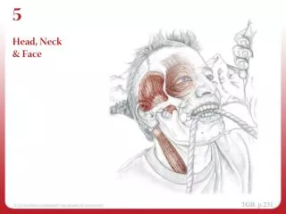
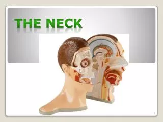
Deep Dissection of the Neck. Chapter 2 The Neck. Section 1 Introduction. Boundaries and Divisions(nape and neck, by the anterior border of the trapezius muscle ) ⅰ . Neck (by the sternocleidomastoid muscle). 1. Sternocleidomastoid region 2. the anterior region 3. the lateral region.

E N D
Deep Dissection of the Neck Chapter 2 The Neck
Section 1 Introduction • Boundaries and Divisions(nape and neck, by the anterior border of the trapezius muscle ) • ⅰ.Neck (by the sternocleidomastoid muscle) 1. Sternocleidomastoid region 2. the anterior region 3. the lateral region ⅱ.Subdivisions of the Triangles. Anterior Triangles ( by hyoid bone)sup. inf. Triangle ( by omohyoid) Carotid Triangle. muscular Triangle Posterior Triangle (by omohyoid ) Occipital Triangle.great supraclavicular fossa
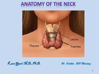
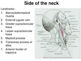
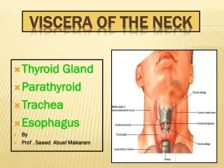
Ⅱ.Surface Anatomy Surface landmarks :1suprasternal (jugular) notch(fossa), 2 hyoid bone, 3 cricoid cartilage, 4 thyroid cartilage, 5 sternocleidomastoid, 6 great supraclavicular fossa
Section 2The Superficialstructures and cervicalFasciaⅠ. The Superficial structures 1.skin 2.superficial fascia(maincontents)1)Platysma, 2)Cutaneous Nervesand thecervical branch of the facial nerve3)Vessels. anterior jugular vein external jugular vein Ⅱ.cervicalFasciadeep cervical fascia.
Ⅱ.Cervical Fascia(deep cervical fascia.) 1.superficial layer2.pretracheal layer(space)3.prevertebral layer(space) 4.carotid sheath
The anterior cervical muscles ⅰ. The suprahyoid muscles (ⅰ) The digastric (ⅱ) The mylohyoid (ⅲ) The stylohyoid (ⅳ) The geniohyoid
The infrahyoid muscles The superahyoid muscles (ⅰ) The sternohyoid (ⅱ) The omohyoid (ⅲ) The sternothyroid (ⅳ) The thyrohyoid
The deep cervical muscles ⅰ. The lateral group of cervical muscles • Scalenus anterior • Scalenus medius • Scalenus posterior Longus capitis Longus colli Scalenus medius ⅱ. The medial group of cervical muscles Scalenus posterior • Longus colli • Longus capitis Scalenus anterior
Erector spinae The muscles of the trunk Ⅰ. The muscles of the back Levator scapulae Rhomboideus Trapezius Ⅰ) Superficial layer ⅰ. Trapezius ⅱ. Latissimus dorsi ⅲ. Levator scapulae ⅳ. Rhomboideus Ⅱ) Deep layer ⅰ. Erector spinae ⅱ. The splenius Latissimus dorsi
The Anterior Triangle of the NeckandSternocleidomastoid region
The Anterior Triangle of the Neckand Sternocleidomastoid region
Axillary artery Internal thoracic artery The subclavian artery Superior thyroid artery Inferior thyroid artery Common carotid artery Transverse cervical artery Thyrocervical trunk Brachial plexus Subclavian artery Suprascapular artery
椎动脉三角: 位于颈长肌、前斜角肌和锁骨下动脉第1段之间。 三角内主要有: 椎动、静脉、甲状腺下动脉、颈交感干、颈胸神经节等。
Vagus nerve. Trace the vagus nerve down to the root of the neck on both sides. Identify the right recurrent laryngeal nerve as it arises from the right vagus and follow it around the subclavian artery to reach the groove between the trachea and the esophagus. Identify the cardiac branches of the vagus nerve.
2.中层——内脏筋膜:位于舌骨下肌群深面,包裹咽、食管颈部、喉、气管颈部、甲状腺和甲状旁腺等器官。2.中层——内脏筋膜:位于舌骨下肌群深面,包裹咽、食管颈部、喉、气管颈部、甲状腺和甲状旁腺等器官。 形成甲状腺鞘,在甲状腺与气管、食管上端邻接处,鞘的后层增厚,形成甲状腺悬韧带。 前下部覆盖于气管者为气管前筋膜; 后上部覆盖于颊肌和咽缩肌者为颊咽筋膜; 包绕颈总动脉、颈外动脉、颈内动、静脉和迷走神经形成颈动脉鞘。
3.深层——椎前筋膜:位于颈深肌浅面, 向上附于颅底,向下续于前纵韧带及胸内筋膜。 两侧覆盖臂丛、颈交感干、膈神经、锁骨下动、静脉。 向下外方,由斜角肌开始,包裹锁骨下动、静脉及臂丛,走向腋腔,形成腋鞘。
(二)颈筋膜间隙 1.胸骨上间隙:封套筋膜在胸骨柄上缘3~4cm处,分深、浅两层,向下附着于胸骨柄前后缘,两层之间为胸骨上间隙。内有颈静脉、颈前静脉下段、胸锁乳突肌胸骨头、淋巴结和脂肪组织等。 2.气管前间隙:位于气管前筋膜和气管颈部之间。内有甲状腺下动、静脉、甲状腺奇静脉丛、头臂干及左头臂静脉。
Back Back 3.咽后间隙:位于椎前筋膜与颊咽筋膜之间,其延伸至咽侧壁外侧的部分为咽旁间隙。 4.椎前间隙:位于脊柱颈部、颈深肌群与椎前筋膜之间。
三、枕三角(肩胛舌骨肌斜方肌三角): (一)境界: 位于胸锁乳突肌后缘、斜方肌前缘和肩胛舌骨肌下腹上缘之间。 (二)内容: 1.副神经:出颈静脉孔后、沿颈内静脉前外侧下行,经二腹肌 后腹深面,胸锁乳突肌上部前缘 穿入并发出分支支配该肌。
At the angle of the mandible, extend the incision backward, below the ear to the mastoid process and then along the superior nuchal line. From the lower end of the midline incision at the suprasternal notch, make an incision along the upper border of the clavicle to the acromion process.
Now carefully reflect the skin flaps laterally to expose the anterior and posterior triangles and the sternocleidomastoid muscle. Avoid damaging the underlying platysma muscle or the supraclavicular and accessory nerves.
If the arm has already been dissected, the skin will have been reflected from the back of the neck; the trapezius muscle will also been reflected laterally.
The superficial fascia is relatively thin and contains little fat; however, it possesses the platysma muscle. Carefully expose the paltysma muscle and note that its fibers arise from the superficial fascia covering the upper part of the pectoralis major muscle.
The fibers form a thin, broad sheet that passes upward and medially across the clavicle, covering the lower anterior part of the posterior triangle and the greater part of the anterior triangle. The muscle is inserted into the lower border of the body of the mandible and the angle of the mouth.
Divide the platysma along the upper border of the clavicle and reflect it upward and forward. Do not damage the underlying supraclavicular nerves and the external jugular vein that lie deep to it.
Locate the three supraclavicular nerves and note that they descend over the clavicle to supply the skin over the thoracic wall down as far as the sternal angle.
Identify the external jugular vein and follow it superiorly to behind the angle of the mandible, where the external jugular is formed by the union of the posterior branch of the retromandibular vein and the posterior auricular vein. Trace the external jugular vein downward until it pierces the deep fascia. This vein varies considerably in size.
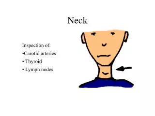
Now clean the sternocleidomastoid muscle and secure the cutaneous branches of the cervical plexus as they pierce the deep fascia at the posterior border of the sternocleidomastoid muscle.
The lesser occipital nerve runs upward along the posterior border of the sternocleidomastoid muscle to be distributed to the skin over the auricle and mastoid process.
The great auricular nerve runs upward and forward to supply the skin over the angle of the mandible and the auricle.
The transverse cutaneous nerve crosses the sternocleidomastoid horizontally to supply the skin over the anterior triangle. The supraclavicular nerves, medial, intermediate, and lateral, descend over the clavicle, where they have already been secured.
Now continue to reflect the platysma as far as the mandible. At the angle of the mandible, examine the deep surface of the platysma and attempt to identify the cervical branch of the facial nerve, which emerges from the lower end of the parotid gland to supply the platysma muscle.
The boundaries of the posterior triangle are the sternocleidomastoid, the trapezius, and the clavicle. The triangle is roofed over by the investing layer of deep cervical fascia.