
Mitigating Cerebral Function Monitor Signal Noise
E N D
Presentation Transcript
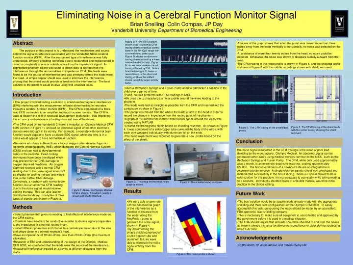
Eliminating Noise in a Cerebral Function Monitor SignalBrian Snelling, Colin Compas, JP DayVanderbilt University Department of Biomedical Engineering Abstract Figure 2: From top to bottom, shown in 2a is a normal CFM tracing characterized by central band in the 10-40µV range with a normal sleep wake cycle. Figure 2b shows an abnormal tracing characterized by a lower, narrow band of activity. Figure 2c shows an otherwise normal tracing affected by EMI. Notice how the tracing in 2c bears a resemblance to the abnormal tracing of 2b as the artifact shows a slightly larger activity band. • Analysis of the graph shows that when the pump was moved more than three inches away from the leads vertically or horizontally, no noise was detected on the CFM. • At a distance of more than twenty inches from the head, no noise could be detected. Otherwise, the noise was shown to dissipate radially outward from the head. • The CFM tracing of the noise profile is shown in Figure 5, and the shielded profile is shown in Figure 6 with the middle recordings shown with shield removed. The purpose of this project is to understand the mechanism and source behind the signal interference associated with the Vanderbilt NICU cerebral function monitor (CFM). After the source and type of interference was fully understood, different shielding techniques were researched and implemented in order to completely minimize outside noise from the impedance signal. An appropriate phantom object was used to obtain data to characterize the interference through the abnormalities in impedance CFM. The leads were found to be the source of interference and was strongest where the leads meet the head. A simple copper shield was used to eliminate the interference, proving that the shield would provide a solution to the interference. The best solution to the problem would involve using well-shielded leads. • Used a Medfusion Syringe and Fusion Pump used to administer a solution to the child over a period of time. • Pump caused problems with CFM readings in NICU. • We used the to characterize a noise profile around the wires leading to the phantom. • The leads were laid as straight as possible from the CFM and inserted into the cantaloupe as show in Figure 3. • The pump was moved from the where the leads attach to the head in order to record the change in impedance from the resting point of the phantom. • A graph of the interference in three dimensional space around the leads was created using MATLAB. • Created electromagnetic shield based on shielding research. As shown in Figure 4, it was comprised of a solid copper tube surround the body of the wires, with each wire wrapped individually with aluminum foil on the ends. • The noise experiment was repeated to generate a new profile based on the effect of the shield. Introduction • This project involved finding a solution to shield electromagnetic interference (EMI) interfering with the measurement of brain abnormalities in neonates through a cerebral function monitor (CFM). The CFM is comprised of a three lead EEG connected to an amplifier and touch-screen monitor. The CFM is used to discern the root of neonatal development dysfunction, thus improving the accuracy and quickness of a diagnosis and overall treatment. • The CFM used by the Vanderbilt Children’s Hospital, an Olympic Medical CFM 6000 (shown in Figure 1), showed an abnormal signal when other electronic devices were brought in its vicinity. For example, a neonate with normal brain function would appear to have a seizure EEG signal, while one who is in a coma would appear to have normal brain function. • Neonates who have suffered from a lack of oxygen often develop hypoxic-ischemic encephalopathy (HIE), which damages the Central Nervous System Figure 6: The CFM tracing of the shield leads with the center tracing showing the shield removed. Figure 5: The CFM tracing of the unshielded profile. Conclusion The noise signal manifested in the CFM tracings is the result of poor lead shielding by the manufacturer, Olympic Medical. An abnormal signal can be generated rather easily using medical devices common to the NICU, such as the Medfusion Syringe and Fusion Pump. The CFM, while only used approximately once a month, is an extremely expensive machine, costing approximately $27,000. The first several hours of a newborn’s life are an integral time in determining brain function. A simple electromagnetic shield was developed and implemented successfully in the NICU setting. While our shield proved to be a valid solution for this problem, it is not adequate to use easily while taking reading on a neonate. Individually shielded leads of a flexible material would be more practical in the clinical setting. (CNS) and can lead to developmental delay in the neonate. Head cooling techniques have been developed which may prevent further CNS damage to oxygen deprived newborns. An oxygen deprived neonate with a normal CFM reading due to the noise signal would not be eligible for cooling therapy and would thus suffer further CNS damage. Conversely, a newborn with normal brain function, but an abnormal CFM reading due to the noise signal, would receive cooling therapy. This can also lead to developmental delay. Examples of three types of signals are shown in Figure 2. Figure 3: The setup for the initial noise graph is shown . Figure 4: The shielded setup is shown . Results Figure 1: Above, an Olympic Medical CFM is shown. A newborn (inset) is shown with leads attached. Future Work • We were able to generate a three-dimensional graph of the interference as a function of distance from the leads, using the MedFusion pump to generate the noise signal, shown in Figure 4. • By implementing the simple shield comprised of a solid copper tube and aluminum foil, we were able to eliminate the noise signal entirely from the CFM. • The best solution would be to acquire leads already made with the appropriate shielding and three wire configuration for the Olympic CFM 6000. To easily accomplish this task, outsourcing the leads should be made by an accredited, FDA approved, lead shielding company. • This is necessary to make sure all equipment in use is tested and approved by the government before it is used in a medical situation. • The FDA should require that all leads should be shielded to and from the device as there is always a chance for device noncompliance or older devices projecting noise over time. Methods • Select phantom that gives no reading to find effects of interference made on the CFM tracing. • Phantom head needs to be conductive in order to show a signal comparable to the impedance of a normal resting infant. • Tested different phantoms and choose to a cantaloupe melon due to the size and shape close to a normal neonate’s head. • Gave an impedance of 10 kilo-Ohms, less than 20 kilo-Ohms (the maximum allowable). • Research of EMI and understanding of the design of the Olympic Medical CFM 6000, we concluded that the leads were the source of the interference. • Measured interference created by a device at different distances from the leads. Acknowledgements Dr. Bill Walsh, Dr. John Wikswo and Steven Steele RN Figure 4: The noise profile is shown.


