Download
1 / 2
20 likes | 187 Views
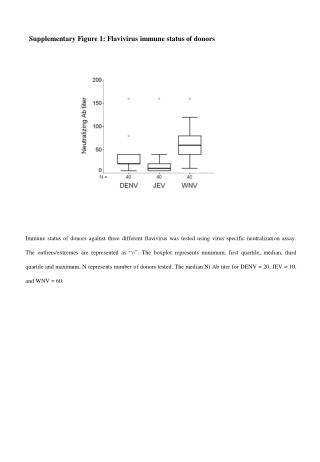
Supplementary Figure 1: Flavivirus immune status of donors.

E N D
Supplementary Figure 1: Flavivirus immune status of donors Immune status of donors against three different flavivirus was tested using virus specific neutralization assay. The outliers/extremes are represented as “ο”. The boxplot represents minimum, first quartile, median, third quartile and maximum. N represents number of donors tested. The median Nt Ab titer for DENV = 20, JEV = 10, and WNV = 60.
Supplementary Figure 2. Total STAT1 levels in JEV infected MDMs STAT1 (91 kD) β-actin (45 kD) Total STAT1 levels in JEV infected MDMs. Primary MDMs were infected with JE057434 and SA14-14-2 strains of JEV. At various time-points (12, 24, and 48 hpi) the cells were harvested , washed thrice in ice-cold PBS. The cells were pelleted and resuspended in loading buffer (Tris-HCl [pH 6.8], SDS, glycerol, 2-mercaptoethanol, 1% bromophenol blue). Cell lysate were then boiled at 95°C for 5 min. Samples were subjected to SDS-PAGE (10%), and the polypeptides were transferred to nitrocellulose membranes. The membrane was blocked for 1 h at RT in Tris-buffered saline containing 5% skim milk powder and 0.1% Tween 20. STAT1 levels were detected using mouse monoclonal anti-human STAT1 (cat. no. 9176; Cell Signaling) as per manufacture’s protocol. Membranes were washed, incubated with horseradish peroxidase- conjugated secondary antibody for 1 h at RT, washed, and finally developed with ECL (cat. no. RPN2232, Amersham Biosciences). β-actin was used as loading control. The figure is representative of two independent experiments. JE057434 SA14-14-2 CC 12 24 48 12 24 48