Download
1 / 67
670 likes | 826 Views
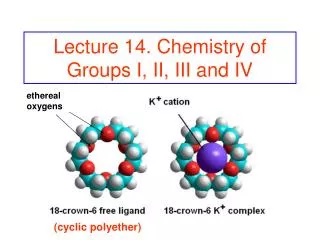
KINE 639 - Dr. Green Section 1 Clinical Physiology I, II, III, IV. Definitions, Concepts, and Hemodynamics. Cardiac Anatomy. Aorta. Superior Vena Cava. Left Pulmonary Artery. Left Atrium. Right Pulmonary Veins. Left Pulmonary Veins. Right CA. Left Anterior Descending CA. Right Atrium.

E N D
KINE 639 - Dr. Green Section 1 Clinical Physiology I, II, III, IV
Cardiac Anatomy Aorta Superior Vena Cava Left Pulmonary Artery Left Atrium Right Pulmonary Veins Left Pulmonary Veins Right CA Left Anterior Descending CA Right Atrium Inferior Vena Cava Left Ventricle Right Ventricle
Cardiac Anatomy Aorta Superior Vena Cava Left Pulmonary Artery Aortic Valve Left Atrium Right Pulmonary Veins Left Pulmonary Veins Right Atrium Tricuspid Valve Mitral Valve Inferior Vena Cava Left Ventricle Right Ventricle Notice that the left ventricle contains more electrically active muscle mass than the right ventricle
Lungs The Normal Heart and Regional Circulation Anterior Cutaway View Pulmonary Semilunar Valve Aorta Left Pulmonary Artery Superior Vena Cava Left Pulmonary Veins Left Atrium Right Pulmonary Artery Aortic Semilunar Valve Bicuspid or Mitral Valve Right Pulmonary Veins Tricuspid Valve Apex Inferior Vena Cava Septum
The Normal Heart - Coronary Artery Anatomy Left Main CA Layers of the Arterial Wall Circumflex Adventitia Media Intima Right CA Marginal Branch Left Anterior Descending CA
Left Ventricular Volumes - Definitions End Diastolic Volume (EDV) Volume at the end of diastole (end of ventricular filling). In a healthy heart this is directly proportional to venous return End Systolic Volume (ESV) Volume at the end of systole (end of ventricular contraction) Stroke Volume (SV) = EDV - ESV Ejection Fraction (EF) = SV EDV NOTE: Resting Ejection Fraction (EF) is the best indicator of both heart performance and heart disease prognosis Left ventricular norm for EF at Rest: approximately 62% Left Ventricular norms for Max Exercise: approximately 80%
Changes in Left Ventricular Volumes with Exercise of Increasing Intensity EDV 120 110 100 90 Left Ventricular Volume (ml) 80 70 SV EDV - ESV = SV 60 50 40 30 20 ESV 10 Rest 300 600 - 750 Peak Exercise Workload or Power (kg meters / min)
Definitions • Cardiac Output: (Q) = HR X SV • Cardiac Index = Q / body surface area • Preload:(EDV) volume of the left ventricle at the end of diastole dependent on venous return & compliance (“stretchability”) of ventricle • Afterload:resistance to ventricular emptying during systole or the amount of pressure the left ventricle must generate to squeeze blood into the aorta. In a a healthy heart this is synonymous with Aortic Pressure &Mean Arterial Pressure (MAP) • Frank Starling Law of the Heart: the heart will contract with greater force as preload (EDV) is increased r more blood in more blood out • Myocardial Contractility: the squeezing contractile force that the heart can develop at a given preload • Regulated by: • Sympathetic nerve activity (most influential) • Catecholamines (epinephrine norepinephrine) • Amount of contractile mass • Drugs
Starlings Law of the Heart and Contractility Starling’s Law: The greater the EDV (blood going in the heart), the more blood comes out of the heart SV (left ventricular performance) uContractility Normal Contractility SV at Preload X - u contractility SV at Preload X – Normal cont. SV at Preload X - d contractility d Contractility (heart failure) The State of Myocardial Contractility determines the amount of blood (SV) that comes out of the heart at a given preload Preload (venous return or EDV) Preload X
Influences on Myocardial Contractility u Contractility related to : Exercise: - ub sympathetic adrenergic nerve output Catecholamines:-Epinephrine & Norepinephrine Excitement or Fear: - Fight or flight mechanism Drugs:- Digitalis & Sympathomimetics d Contractility related to: Loss of contractile mass: - Most likely due to heart attack Myocardial muscle disease: - Cardiomyopathy Drugs: - Anesthetics, Barbiturates
Definitions • Arteriovenous Oxygen Difference (AVO2D) the difference in oxygen content between arterial and venous blood • measured in ml% - ml O2 / 100 ml blood • Oxygen Consumption (VO2) - the rate at which oxygen can be used in energy production and metabolism • “absolute” measures: L O2 / min , ml O2 / min • “relative” measures: ml O2 / kg body wt. / min • Fick equation: VO2= Q X AVO2D • Maximum Oxygen Consumption (VO2max) maximum rate at which a person can take in and utilize oxygen to create usable energy • defined as plateau of consumption rate increase • often estimated with VO2peak • Myocardial Oxygen ConsumptionVO2 of the heart muscle (myocardium) • "estimated" by RPP: HR X SBP
Definitions • Systolic Blood Pressure (SBP) pressure measured in brachial artery during systole (ventricular emptying and ventricular contraction period) • Diastolic Blood Pressure (DBP) pressure measured in brachial artery during diastole (ventricular filling and ventricular relaxation) • Mean Arterial Pressure (MAP) "average" pressure throughout the cardiac cycle against the walls of the proximal systemic arteries (aorta) • estimated as: .33(SBP - DBP) + DBP • Total Peripheral Resistance (TPR)- the sum of all forces that oppose blood flow • Length of vasculature (L) • Blood viscosity (V) • Vessel radius (r) TPR = ( 8 ) ( V ) ( L ) ( p ) ( r 4 )
Cardiovascular Hemodynamic Basics Pressure (MAP) P aorta – P vena cava = = Resistance (TPR) (8) (V) (L) () (r 4) Flow (Q) = () (Pa – Pv) (r 4) (8) (V) (L) Flow (Q) Normally Resting Q is about 5 - 6 liters / minute V = viscosity of fluid (blood) flowing through the pipe L = length of pipe (blood vessel) r = radius of the pipe (blood vessel) Pa = aortic pressure Pv = venous pressure
Respiratory Physiology - Definitions • Minute Ventilation (VE)– amount of air passing through the lungs in one minute • Dyspnea- breathing difficulty • Respiratory Exchange Ratio - amount of CO2 expired by the lungs divided by the amount of O2 extracted from the air in the lungs (VCO2 /VO2 ). RER = .7 r 100% fat 0% carb RER = .85 r 50% fat 50% carb RER = 1.0 r 0% fat 100% carb
Neurophysiology - Definitions • Afferent - sensory nerves - going toward spinal column • Efferent - effector nerves - going away from spinal column Motor cortex (control of voluntary muscles) Central sulcus Basil nuclei (planning, organizing, coordinating motor movements) Sensory cortex (Sensation & pain) Essential Knowledge of the Areas of the Brain in Green Sulcus Corpus callosum Gyrus Choroid Plexus Broca’s area (motor, speech Taste area Thalamus Parietal lobe General interpretive area Parietal lobe Frontal lobe Pineal gland Frontal lobe Occipital lobe Occipital lobe Visual area Reticular activation system (sleep wake) Hypothalamus Temporal lobe Pituitary gland Optic chiasma Cerebellum (movement coordination & balance) Lateral sulcus Cerebellum Pons Brainstem (control of HR, BP, and respiration) Medulla oblongata Auditory cortex Vestibular area Temporal lobe Fourth Ventricle Central canal
Organization of the Nervous System Nervous System Central Nervous System (CNS) Peripheral Nervous System (PNS) Spinal Cord Brain Motor (efferent) Neurons Sensor (afferent) Neurons Autonomic Nervous System Somatic Nervous System Voluntary movement Output to Skeletal muscles Involuntary (Reflexes) Input from Internal receptors Output to smooth muscles and CVS Sympathetic Motor System Parasympathetic Motor System Relaxing responses Neurotransmitter: acetylcholine Cholinegeric nervous system Flight or Fight responses Neurotransmitter: nonadrenaline Adrenergic nervous system
Adrenergic Receptors & Associated Responses • a1stimulation: • Constriction of blood vessels • Vascular smooth muscle activation • Constriction of lung bronchioles • Constriction of bladder muscles • u myocardial cardiac contractility • Relaxation of GI tract • a2stimulation: • u central sympathetic outflow • u release of E & NE • a1 & b1 receptor activation • Constriction of lung bronchioles • b1stimulation: • u in HR • u in myocardial contractility • u in Renin secretion • u fluid retention • b2stimulation: • Dilation of lung bronchioles • Dilation of blood vessels Agonist – body molecule or drug “stimulator” Antagonist - body molecule or drug “in-activator” a2Agonists b1 & b2Agonists Agonists in the adrenergic system are primarilyepinephrineandnorepinephrine Antagonists are many times associated with drugs known as “blockers” i.e.“b-blocker”or“a-blocker” Responses
Brain Lungs Arteries (Stiff Inflexible “Pipes”) Veins (Flexible Compliant “Pipes”) Intima Intima Valve Elastin Elastin Media Media Liver Stomach Externa Externa Pancreas Serosa Intestines Kidneys The Systemic Circulation Arterioles and Pre-capillary Sphincters Skin Muscle
Microcirculatory Anatomy – a Capillary Bed Arteriole Smooth Muscle Pre-capillary Sphincters (closed in this illustration) Anastomosis (Shunt) Capillary Bed Metarteriole Capillary in Cardiac Muscle (arrows) Venule
Development of the Driving Pressure in the Human Cardiovascular System 100 Arterial 100 Pressure Normal Resting Pressure Driving the Blood from Left Ventricle to Vena Cava: 100 - 0 = 100 mmHg (mm Hg) 26 Mean Circulatory Filling Pressure 7 7 0 7 6 7 0 Central 0 Venous Pressure (mm Hg) 1 5 0 Normal Resting Cardiac Output Cardiac Output (Q) (Liters / min)
Arterial Pressures in Maroon The “Closed” Cardiovascular Hemodynamic System Venous Pressures in Blue LV PO2 = 160 PCO2 = .3 RV LA RA LUNGS AORTA (13) (100) (3) (0) PO2 = 100 PCO2 = 40 9% of blood volume (7) SYSTEMIC ARTERIES Ohms Law: Flow (Q) = upstream pressure – downstream pressure resistance (92) low compliance 13% of blood volume VEINS (CAPACITANCE VESSELS) (20) high compliance 64% of blood volume (40) (2) CAPILLARYBEDS ARTERIOLES PO2 = 40 PCO2 = 46 7% of blood volume Systemic Circulation = 100 mmHg – 0 mmHg = 100 ml / sec = 6 liters / min Flow (Q) 1 mmHg sec / ml
Pressure, Flow, and Resistance by Vascular Cross Sectional Area Veins Vena Cava Veins Vena Cava Capillaries Venules Capillaries Venules Arterioles Arterioles Arteries Arteries Aorta Aorta Cross Sectional Area 100 mmHg Flow Velocity 0 mmHg 100 mmHg Relative Resistance to Flow Mean Arterial Pressure 0 mmHg
MV is an Atrio-Ventricular Valve (AV) and is bi-cuspid Aortic valve is a semilunar valve The Cardiac Cycle Aortic Valve opens Aortic Valve closes SYSTOLE 120 Aortic Pressure (mmHg) 80 Left Ventricular Pressure (mmHg) MV closes MV opens Atrial Contraction Passive Ventricular Filling Left Atrial Pressure (mmHg) 0 120 Left Ventricular Volume (ml) 0 0 .2 Time (sec) http://www.youtube.com/watch?v=yGlFBzaTuoI&feature=related http://www.youtube.com/watch?v=dYgYcH7R29I&NR=1
Using Ventricular Pressure Curves as Indices of Contractility & Cardiac Function dP/dt = change in pressure per unit of time dP/dt dP/dt Normal Heart Failure 120 Pressure Note elevation in end diastolic pressure indicating the build up of pressure in the heart due to failure 0 Time
Cardiac & Vascular Function Curves Cardiac Function Curve - illustrates Q what happens to Q when Pv changes. 5 CARDIAC OUTPUT (Liters / min) operating point for the cardiovascular system 2 Pv CENTRAL VENOUS PRESSURE (mmHg) CARDIAC PRELOAD (mmHg) RIGHT ATRIAL PRESSURE (mmHg)
Cardiac Function Curve - Q is the dependent variable (in effect, Q is controlled by venous return & TPR) Sympathetic Stimulation ( Exercise) Normal Curve CARDIAC u Intrapleural Pressure ( Pressure in the chest cavity ) To maintain same Q, CVP must u OUTPUT (Liters / min) 5 u TPR r u afterload Parasympathetic Stimulation or Heart Failure 2 7 CENTRAL VENOUS PRESSURE (mmHg) ( can also be thought of as CARDIAC PRELOAD or RIGHT ATRIAL PRESSURE )
Cardiac & Vascular Function Curves Cardiac Function Curve - illustrates Q what happens to Q when Pv (venous 5 return - preload) changes. CARDIAC OUTPUT operating point for the cardiovascular system (Liters / min) Vascular Function Curve - illustrates what happens to Pv when Q changes. 2 7 Pv CENTRAL VENOUS PRESSURE (mmHg) CARDIAC PRELOAD (mmHg) RIGHT ATRIAL PRESSURE (mmHg)
Vascular Function Curve - central venous pressure is dependent variable (in effect, CVP and venous return are controlled by Q) Sympathetic Stimulation Exercise r venoconstriction r u total active vascular volume ( Transfusion: u blood Volume ) CARDIAC OUTPUT Normal Curve (Liters / min) 5 Decreased Arteriolar Resistance: d TPR ( Exercise ) ( Peripheral Vasodilation ) Increased Arteriolar Resistance ( Peripheral Vasoconstriction) (u TPR rd venous return r d CVP) 2 7 CENTRAL VENOUS PRESSURE (mmHg) can also be called CARDIAC PRELOAD
Changes in Cardiac & Vascular Function Curves with Exercise • During Exercise, an u in sympathetic output will cause: • 1. venoconstriction (VF curve shifted rightward) • 2. d TPR r vasodilation (VF curve rotated upward) • (caused primarily by vasodilator metabolites) • 3. u HR & contractility (cardiac curve shifted upward) 8 - 13 CARDIAC OUTPUT (Liters / min) 5 7 2 11 CENTRAL VENOUS PRESSURE (mmHg) can also be called CARDIAC PRELOAD
Changes in Cardiac & Vascular Function Curves with Acute Compensated Heart Failure CARDIAC OUTPUT 1 3 (Liters / min) Q = 5 Liters / min 2 CENTRAL VENOUS PRESSURE (mmHg) can also be called CARDIAC PRELOAD 1. Normal point of operating system & normal cardiac output 2 Pump begins to fail r Q falls below normal resting levels 3. Renin-angiotensin system activated r fluid retained r u MCFP r u Q
Changes in Cardiac & Vascular Function Curves with De-compensated Heart Failure CARDIAC OUTPUT 1 3 Q = 5 Liters / min (Liters / min) 4 2 5 Cause of peripheral edema CENTRAL VENOUS PRESSURE (mmHg) can also be called CARDIAC PRELOAD • 1. Normal point of operating system & normal cardiac output • 2. Pump begins to fail r Q falls below normal resting levels • Renin-angiotensin system activated r fluid retained r u MCFP r u Q • Pump decline continues and Q falls once again • More fluid is retained to try and compensate, but now Q is below a level where normal fluid balances can be maintained r r pattern continues
Left Ventricular Pressure Volume Loop Aortic Valve Closes ESV ESP Aortic Valve Opens 120 Left Ventricular Pressure (mmHg) SV Isovolumic contraction Mitral Valve Closes EDV EDP 6 Slope of dashed line: ventricular contractility 40 140 Mitral Valve Opens Ventricular Filling Begins Volume (ml)
Effects of an Increase in Preload on Left Ventricular Pressure Volume Loop u Ejection Pressure 120 Left Ventricular Pressure (mmHg) u SV u EDV u EDP 6 40 140 Volume (ml)
Effects of an Increase in Afterload on Left Ventricular Pressure Volume Loop u ESV u ESP 120 Left Ventricular Pressure (mmHg) d SV 6 40 140 Volume (ml)
Mechanism of Control of Cardiovascular and Respiratory Systems
atrial stretch / pressure receptors located in right & left atria, junction of right atria and vena cava, and junction of left atria and pulmonary veins Sites of Cardiorespiratory Control
Cardiorespiratory Control • Heart Rate– Neurohormone (neurotransmitter) and CNS (medulla) regulation • Parasympathetic vagus control (Neurotransmitter: Acetylcholine) • Vagal control is dominant at rest – influence is withdrawn when exercise begins • Sympathetic cardioacceleration (Neurotransmitters: EPINEPHRINE & NOREPINEPHRINE) • Baroreceptor influences • Sympathetic discharge indirectly proportional to firing rate • Parasympathetic discharge is directly proportional to firing rate • dpressure r dreceptor firing r usympatheticsr u HR r u pressure • u pressure r ureceptor firing r uparasympatheticsr d HR r d pressure • Atrial Stretch receptors: u receptor stretch ru ANP ruNa+excretionru urine output • d receptor stretch ruADHrdNa+excretionrd urine output • AtrialNatriureticPeptide released by myocytes in the atria r u urine flow r d BP • Aniti-Diuretic-Hormone (vasopressin) released by pituitary r d urine flow r u BP • Chemoreceptor influences • Main function: protect brain from poor perfusion • u O2or d CO2r uparasympathetic discharge r d HR • dO2 or u CO2r dpH rpressor area stimulation in medulla r uHR ADH Molecule
Cardiorespiratory Control • Stroke Volume (SV) – regulated by Frank Starling mechanism • u venous returnr u EDV r u stroke volume • Cardiac Output (Q)– main determinant: body O2 needs • Autoregulated by two distinct mechanisms • Intrinsic changes in preload, afterload, and SV • u afterload r initial d in Q r u EDV (preload) r uSV back to normal • Extrinsic hormonal influences • Norepinephrine release r u HR and SV
Cardiorespiratory Control • Blood Pressure– influenced by 4 major factors (some interrelated) • Total peripheral resistance • Baroreceptor (BR) and CNS Influences • u BP r u BR firing rate r vasodilation r d BP • d BP r d BR firing rate r u sympathetics r u BP • Chemoreceptor influences • dO2,u CO2, d pH r CNS stim. r vasoconstriction • Circulating catecholamine influences • E and NE have varying effects on TPR • E and NE usually activate areceptors r u TPR • Fight or flight response • Q • Blood Volume • Renin – Angiotensin System
HYPOTENSION HYPOVOLEMIA Renin - Angiotensin System u H2O reabsorbed d renal profusion u sympathetic tone u ADH (vasopressin) d stretch in afferent arteriolar JG cells d NACL delivery to macula densa cells u messangial cell contraction uRENIN d GFR uangiotensin I d stretch receptor activation in atria, aorta, and carotid sinuses uangiotensin II u BLOOD PRESSURE via vasoconstriction (angiotensin II is a potent vasoconstrictor) u aldosterone neg feedback u Na+ re-absorption (and K+ excretion) Controls Body Fluid Balance and Associated Regulation Mechanisms and Pathways u thirst (thirst is more strongly regulated by osmotic receptors in the hypothalamus) u ECF volume ru BP neg feedback dRENIN
Dehydration • Dehydration: the loss of body water and associated electrolytes • Causes: • Gastroenteritis (viral / bacterial infection r vomiting & diarrhea) - most common • Diseases: yellow fever, cholera, • Excessive alcohol consumption • The excess fluid is flushed out by the kidneys: u water usage rdehydration • Most liquors have congenerswhich are toxic to body r removal necessary • The clearer & better quality your liquor (vodka & gin) the less congeners • more distillation cycles r better quality • When you drink, head vessels dilate….constriction next morning r headache • Congener removal done by liver: d liver glucose r hypoglycemia & lethargy • Prolonged exercise without fluid replacement (heat exhaustion & heat stroke risk) • Diabetes: hyperglycemia r u glucose excretion r u water loss r dehydration • Shock: blood loss due to some hypotensive state caused by injury or disease • Gastrointestinal blood loss: bleeding from ulcers or colorectal cancer
Dehydration • Signs & Symptoms of dehydration: • Dry mouth, dry swollen tongue, rapid heart rate (possible chest palpitations) • Lethargy (sluggishness), confusion • Poor skin turgor (a pinch of skin does not spring back into position) • Good test for ailing elderly folks • Elevated BUN (renal function test): NH4 metabolized in liver & excreted by kidneys • Elevated creatinine r d GFR (kidney clearance of waste products) • Increased blood viscosity • Headache • Fluid loss r low blood pressure r dizziness upon standing up • A high urinary specific gravity (comparison of density to water: 1 gram / cm 2) • Treating Dehydration • Sip small amounts of water • Drink carbohydrate / electrolyte solutions: Gatorade, Pedialyte, etc. • If core body temperature > 104 0 + d BP or u HR r consider IV fluid replacement
Cardiorespiratory Control • Skeletal Muscle Blood Flow – autoregulated – 2mechanisms • Mechanism 1: Vasodilator Metabolites • Usually overrides adrenergic neurohormone control • Mediated by vasodilator metabolite (VDM) buildup & removal • Adenosine (ATP by-product), CO2, H+, prostaglandins • Exercise Example – (negative feedback control) • Muscle exercises r VDM’s released r u vasodilation • u vasodilation u blood flow r VDM’s removed r vasoconstriction • Mechanism 2: Myogenic response • Involves stretch activated Ca++ channels (negative feedback control) • u blood flow r vessel stretch r Ca++ channel activation • u [Ca++ ] in smooth muscle r vasoconstriction r d flow
Cardiorespiratory Control • Systemic Blood Flow During Exercise: • Autonomic influences • Sympathetic outflow & circulating catecholamines • a activation r vasoconstriction in non - exercising tissue • Redistribution of blood flow during maximal exercise • - NC in brain blood flow - 500 ml/min u to heart • - 11,300 ml/min u to muscle - 400 ml/min u to skin • - 500 ml/min d to kidneys - 800 ml/min d to viscera • - 200 ml/min d to various other parts of the body
Cardiorespiratory Control • Respiration: Minute Ventilation (VE) = Tidal Volume X Respiratory Rate • GenerallyControlled via central chemoreceptors in the medulla-pons respiratory center • Peripheral chemoreceptors • u blood CO2 content r receptor activation ru VE • dblood O2 content r receptor activation ru VE • Central chemoreceptors in the medulla respiratory center – Dominant Influence • u blood CO2 & lactate r receptor activation ruVE • PaCO2 ru HCO3¯ + H+ r H+ activates receptor ru VE • Respiratory control during exercise – no consensus but research suggests: • Muscle spindle & proprioceptor activation ru VE at early onset of an exercise bout • Respiratory centers (medulla) sends afferent signals to expiratory muscles during exercise • u venous returnr atrial receptor activation ru VE ?? • Intrapulmonary receptor activation ru VE ?? • Peripheral chemoreceptors may play a role in steady state & high intensity exercise VE ?? • Minute ventilation mechanistic changes during an u in exercise intensity • Low exercise intensity: VEu by both u TV and u RR • High exercise intensity: VE u by u RR only • Notes: • O2 cost of breathing during exercise: 4.5% VO2 (low int.) up to 12.5%VO2 (high int.)
Acute Responses to Aerobic Exercise • Oxygen Consumption (VO2) • u VO2 in direct proportion to u workload (power requirement of exercise) • Expressed in both relative and absolute terms • Relative: ml O2/kg/min Absolute: ml/min or L/min • Average VO2max for 40 year old male 37 ml/kg/min • Oxygen consumption linked to caloric expenditure (1 liter of O2 consumed = 5 kcal) • Heart Rate • u up to 3 times resting value at peak exercise (mainly due to dtime spent in diastole) 180 160 140 100 Heart Rate • HR – VO2 relationship is linear until about 90% VO2max 1.0 2.0 3.0 Oxygen Uptake (L / min) 50 150 250 Workloads (Watts)
Acute Responses to Aerobic Exercise • Stroke Volume • u up to 1.5 resting value at peak exercise • Increase levels off at 40% - 50% VO2 max?? • u in venous return r u EDV (Starling mechanism) • d ESV eluding to an u in myocardial contractility • u ejection fraction rest: 58% max exercise: 83% • Cardiac Output (Q) • u up to 4 times resting value at peak exercise (u is rapid at onset, then levels off) • u Q r u venous return • Venous return mediated by and related to: • Sympathetic venoconstriction • Muscle pump • u inspiration r d thoracic pressure • Blood flows to an area of reduced pressure • u inspiration r u abdominal pressure • Contraction of abdominal muscles r squeezing of abdominal veins ?? 120 110 70 120 110 70 Stroke Volume (ml/beat) Recent Findings All Older Studies 25% 50% 75% 100% 25% 50% 75% Percentage of VO2 max Percentage of VO2 max
Arteriovenous oxygen difference • Difference in [O2] between arterial and mixed venous blood • Illustrated by the oxyhemoglobin desaturation curve • u approximately 3 fold from rest to max exercise • At rest, about 25% of arterial O2 is extracted • At peak exercise about 75% - 85% of arterial O2 is extracted • Blood Pressures and Resistance to Flow • SBP: u - failure to u signifies heart failure • DBP: slight u or slight d or NC • MAP: slight u • TPR: d - mainly due to vasodilation in exercising muscle • Coronary (Myocardial) Blood Flow • 4.5% of Q goes to myocardium at rest and at peak exercise • This increase is due to u MAP and CA vasodilation • Blood Flow to the Skin • u as exercise duration u to allow for heat dissipation • d at max exercise to meet exercising muscle demands • u during exercise recovery, again for heat dissipation Acute Responses to Aerobic Exercise