Download
1 / 16
180 likes | 373 Views
Cellular and Colonial Morphology Aseptic Technique, and Streak Plate. Labs 1 and 2. Applications of Light Microscopy. Observe less detailed features of intact cell than advanced microscopy (i.e. electron and laser)

E N D
Cellular and Colonial MorphologyAseptic Technique, and Streak Plate Labs 1 and 2
Applications of Light Microscopy • Observe less detailed features of intact cell than advanced microscopy (i.e. electron and laser) • Shape, presence of flagella, diagnostic stain, arrangement of cells, some large internal features • <1000X magnification • Brightfield • Stained cells or cells with color/contrast • External features • Phase contrast • Live transparent (unstained) cells • Some internal features • Dark-field • Live transparent cells • Greater resolution; more features; internal and external
Path of Light: Brightfield • Illuminator • Filter for shorter λ; blue light • Condenser • Focuses light into specimen • Diaphragm (iris) • Controls amount of light to specimen • Specimen on slide • Diffracts light as it passes throughimage produced • Objective lenses at nosepiece • Magnifies image 10, 40, or 100 times • Image inverted • Head with prism • Direct light path into ocular lens • Ocular lens magnifies image 10X • Image path sent to eye
Magnification Versus Resolution • Magnification = increase in apparent size • Objective and ocular lenses • Resolution = clarity • Ability to see 2 nearby objects as distinct objects • Resolving power depends on numerical aperture of lens • Ability to gather light; increase aperture and increase resolution of lens • Increase magnification must increase aperture; aperture limit • Highest resolution possible is 0.2μm with 100X lens • Steps you can take to increase resolution • GET MORE LIGHT TO LENS! • Oil immersion with 100X lens • Same refractive index as glass; prevents loss of light to air • Small wavelength light (blue light) • Adjust diaphragm and condenser as you increase magnification • Course focus and fine focus to focus image • Clean lenses and slide
Microscopy • Field of vision • Smaller at higher powers; must CENTER object or you will lose it at higher power • Mechanical stage adjuster!!! • Parfocal • Focus under low power then move to high power immediately; don’t move focus in between; then fine focus after you get to high power. • Contrast versus resolution. • you need light to increase resolution, but too much light can decrease contrast; must find a happy medium. • What happens if I go from low power to high power and the cells disappear? • Field of vision narrowed—center specimen • Not enough light—open diaphragm • Slightly out of focus—use fine focus • Resolution not optimal—clean lenses or slide, use oil if using 100X lens, open diaphragm, • The lens is not clicked all the way in place—make sure it clicks
Cellular Morphology • Prokaryotes • Rods (strep or single arrangement) • Cocci (staph, strep or single arrangement) • Spirillum • No nuclei present. • Must have cells under oil immersion to see any detail. • Eukaryotes • Cells larger than prokaryotes • Can’t always see nucleus • Fungi • Molds versus single celled fungi • Yeast • Candida albicans; vaginal yeast infection and thrush • Saccharomyces cerevisiae; bakers yeast; brewing yeast
Cellular Morphology Continued • More Eukaryotes • Fungi • Molds • Filamentous due to hyphae; multicellular • Penicillium • Yes it makes penicillin; grows on bread • Aspergillus, • Some produce aflotoxins (pathogenic) • Rhizopus • Classic bread mold and house mold • Yeast • Saccharomyces (bread and beer yeast) or Candida albicans (yeast infections) • Algae • Have pigments associated with them; single cell • Dinoflagellate called Peridinium • Protozoa • Single cell • Trypanosoma gambiensae • Causes Sleeping Sickness; nervous disorders; central Africa; tsetse flies • Helminth • Multi cell • Schistosoma mansonni • Causes schistosomiasis
Microbes in the Environment • Where do microbes live and where do they not live? • Sterile media versus culture. • Autoclave • To inoculate is to purposely grow an organism by putting it in some media, and to contaminate to to accidentally grow an unwanted organism. • Broth versus solid media • Agar • Slant versus plate media • Incubation
Growth • Growth in brothcloudy or turbid • Pellicle, sediment, flocculant • Growth on platescolonial morphology • Colony and colonial morphology • Margin, elevation, color and colony shape • Page 52 for terms to use • Observe with dissecting scope • Dissecting scopes used to observe 3D structure of larger objects while microscopes used to see 2D surface of smaller objects.
Describing Cultures Terms for broth growth Terms for colonial morphology
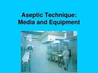
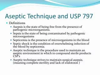
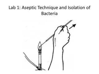
Aseptic Technique • Types of media • Deep • Slant • Broth • Sterile • Asepsis • Inoculate • Demo the technique • Describing your cultures: • Motile versus non-motile • Slant descriptions • Broth descriptions (pellicle, flocculant, sediment, turbid) • Can you tell if a culture is contaminated? How?
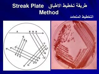
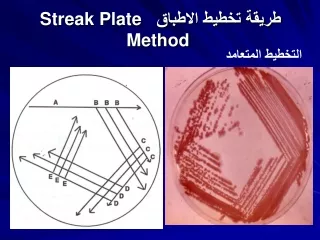
Streak Plate • Streak for isolated colonies • Pure culture • All cells came from same parental cell • Genetic information is identical for most part • Isolated colony • Streak plate used to get isolated colonies pure culture