Download
1 / 54
550 likes | 795 Views
Explore the intricate stages of facial and palate development, from formation of the pharyngeal arches to palate fusion. Understand the critical periods and anomalies involved in facial development.

E N D
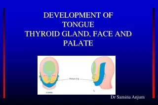
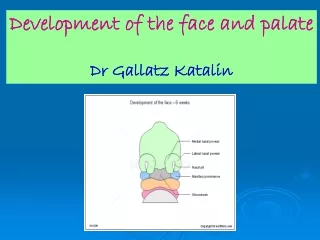
Development of the face and palate Dr Gallatz Katalin
Development of the face and pharyngeal arches 4-5.week Pharyngealarchesbegintodevelop inthe 4th weekastheneural crestcellsmigrateintothefuture head and neckregion.
The pharyngeal (branchial) apparatusconsist of: - pharyngealarches - pharyngealpouches - pharyngealgrooves - pharyngealmebranes
Each pharyngeal arch contain: - cartilage - nerve - artery - myoblast - mesenchyme
Stomodeum stomodeum: a wide shallow depression in the face, limited in its depth by the buccopharyngeal membrane
STOMODEUM SURROUNDED bythefacialprominences Frontalprominence (frontonasal) fromthemesenchyme coveringtheforebrain Maxillaryprominence frommesenchyme of the I. pharyngealarch Mandibularprominence Fromthemesenchyme of the I. pharyngealarch
4.week Onbothsides of thefrontonasalprominencesthickeningsofsurfaceectodermformthe NASAL PLACODS
5. week 1.The nasal placods invaginate to form the nasal pits 2.Medial and lateral nasal processesare formed around the nasal pits 3.Maxillary and lateral nasal processes are separated by the nasolacrimal groove nasolacrimal duct
NASAL PITS MEDIAL AND LATERAL NASAL PROMINENCES
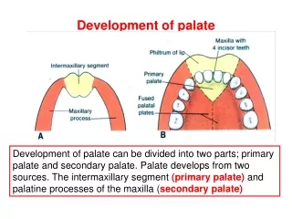
7-10. week Maxillary prominences grow and press the medial nasal processes, they unite and fuse with the maxillary prominences. The inferior part ot fused medial nasal prominences form the INTERMAXILLARY SEGMENT
Intermaxillary segment is composed of 1. labial component philtrum of the lip 2. upper jaw component alveolar process of the incisor teeth 3. palatine component primary palate
- The ectodermal thickening betwen the lateral nasal and maxillary invaginates into the underlying mesoderm, the nasolacrimal groove and cord is formed, later it canalicularises and it becomes NASOLACRIMAL DUCT. - The maxillary prominences unite with lateral nasal processes and also with the mandibular prominences.
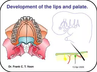
Palatogenesis 5-12th week Criticalperiod 6-9th week The palatedevelopsfromtwo primordia: - theprimarypalate - thesecondarypalate
Development of the palate Primarypalate developsfromthe palatineportion of the intermaxillarysegment Secondarypalate developsfromthe palatineprocess of themaxillaryprominence
- Palatineprocesses(orpalatalshelves) project inferolaterallyoneachside of thetongue. - Asthemandibledevelopthetonguemovesinferiorly, thepalaineprocesseselongate and ascendtothehorizontalposition. Theyfuseinthemedianplane and alsofusewiththenasalseptumandwith theprimarypalate Nasalseptumdevelopsas a downgrowthfromtheinternal part of themerged medialnasalprominences
Derivates of the facial prominences Frontonasal prominence frontal part: forehead, root of the nose medialnasal process: - dorsum and tip of the nose, nasal septum intermaxillary segmentum: philtrum, alveolar process of the incisor teeth, primary palate lateral nasal process: alae (wings) of the nose Maxillary prominences: maxilla, lateral part of the upper lip, upper part of the cheek secondary palate Mandibular prominence: mandible, lower lip, inferior part of the cheek
Development of the nasal cavity - oronasalmembrane - primitivechoana • primary and secundary palate - nasalconchas - olfactoryregion - paranasalsinuses
Summary of the development of the face 1. Development of the stomodeum and the facial prominences, 2. appearence of the nasal placods nasal pits 3. development of the medial and lateral nasal prominences 5.week 4. growth of the maxillary prominences, 5. fusion of the medial nasal processes, 6. formation of the intermaxillary segment, 7. development ot the primary palate 6-7. week, 8. formation of the nasolacrimal sulcus and duct, 9. fusion of the maxillary prominence with the and medial and lateral nasal prominences, 10. secondary palate, separation of the nasal and oral cavity
Development of the Face MOVIE v
GENES IN DEVELOPMENT OF THE FACE
Malformations Median cleft lip Lateral cleft lip (cheiloschisis) Cleft of the upper jaw (gnatoschisis) Cleft palate (palatoschisis) Oblique facial cleft Transverse facial cleft Macrostomy, microstomy Cleft uvula
Malformations Median cleft Cleft of inferior lip Obligue facial cleft Macrostomia Microstomia Bifid nose and incomplete median cleft
Congenital anomalies of the face 1.Lateral cleft lip ( hare lip): Due to failure of fusion between the maxillary process with the intermaxillary segment, unilateral or bilateral .
Congenital anomalies of the face 2. Median cleft lip : • Due to incomplete union of the 2 medial nasal process in the midline .
3. Oblique facial cleft : Due to failure of union of the maxillary processes with lateral nasal processes.
Development of the tongue ANTERIOR 2/3 At the end of the 4th week on the base of the primordiali pharynx 3 tubercule develops from the mesenchymeof the Ist pharyngeal arch, The pairedTUBERCULUM LATERALEand between and behind them the TUBERCULUM IMPAR. The lateral tubercules develop quickly, overgrow the tuberculum impar and unite in the midline ( median lingual sulcus)
Development of the tongue POSTERIOR 1/3 From the II-III. pharyngeal arch mesenchyme. COPULA from the the ventromedial part of II. pharyngeal arch HYPOBRANCHIAL EMINENCE from the ventromedial part of III-IV. pharyngeal arch
Development of the tongue EPITHELIUM Anterior 2/3 ectoderm Posterior 1/3 endoderma MUSCLES From the myotomes of the occipital somites
DEVELOPMENTAL MALFORMATIONS BIFID TONGUE