
Abstract
Dual Light Controlled A rabinose B iosensor.

Abstract
E N D
Presentation Transcript
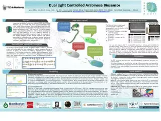
Dual Light Controlled Arabinose Biosensor Aguilar, Mónica; Cano, Nelson ; Colunga, Indira I. ; Díaz, Aldo A. ; Guerrero, Israel ; Machado, Rodrigo ; Maycotte, David ; Morales, Cintli C. ; Nieto, Mariana ; Taveras, Rossel ; Vásquez Jorge A. ; Villarreal, Antonio. MishraPrashant K. (pmishra@itesm.mx); Vázquez-Flores, Sonia (svazquef@itesm.mx ) Abstract Howdoesitwork? Results and discussion PCR Reaction of the BackboneBBAJ_04450 Integrating the work of many other previous iGEM teams (Tokyo NoKoGen 2010, Chiba 2009, 2010, British Columbia 2009, Cambridge 2010, UNAM-Genomics México 2010, ITESM Monterrey 2010), the aim of this project is to develop a way of giving a cell the command to perform a function at user’s will, improving current lock-and-key designs. A novel mechanism based on an E. coli chassis, was designed with two main objectives: to sense arabinose reporting its concentration and to use light receptors to trigger the expression of the required pathways. The first receptor enables E. coli activity, expressing the arabinose sensing mechanism; whereas the second receptor activates a quick deactivation (degradation) of the sensing mechanism, depriving the cell of that capability. The following composite parts were cloned into DH5α and BW27783 competent cells using the standard CaCl2 transformation protocol: Constitutive promoter + Crx-st+ RBS + GFP + Terminator Constitutive promoter + Ta-st+ Terminator Constitutive promoter + iTa-st+ Terminator a + b + c 1 % gel, V Ligatedpieceswithlambda phage T4, 4µl of Fermentas O’GeneRulerTM1kb ; digested DNA 10 µl in 0. 5µl of 6x Orange DNA Loading Dye . The gel was run at 100 V for 30 minutes. 2% gel with PCR product of theconstructedBackbone BBAJ_04450 with a 4µl of Fermentas O’GeneRulerTM 100 kb. Assembly and construct description Conclusions We have some evidence that the construct fully assembled. Bacteria were inoculated into several Petri dishes with LB agar and the appropriate antibiotic (Chloramphenicol) to screen for transformants. After a 24 hour incubation at 37°C, there was growth in the dishes that had bacteria transformed with all the composite parts. These dishes were submitted to UV light to see if they produced fluorescence, although there was evidence of reaction, it was not conclusive, nor uniform in all the cultures. The levels of GFP expression might not have been high enough to directly observe fluorescence. The construct combining three plasmids, the green receptor activates the expression of the recA final product, this RecA protein binds into the operators, allowing the expression of pBAD´s, in presence of arabinose, depending on the arabinose concentration; if there is a high concentration, the low concentration plasmid will be inactivated by the iTa-st. If there is a low concentration, the plasmid will activate the pBAD´s and along with keys and anti-keys to avoid the expression of both the fluorescent proteins. The GFP fluorescent mechanism was successfully designed, incorporated and tested in a bacterial system. The system has yet to be tested for low and high arabinose quantities, and quantify the lowest detection level of fluorescence in a fluorometer. Nonetheless, this study opens a new window for further experimentation for concentration dependent detection mechanisms for other metabolites. Fig. 1. Plasmid assembly Fig. 2. Construct conformation and part assembly Future research Modelling Millions of numbers. There are a wide variety of biosensors in the World, responsible for detecting a specific factors but not all of them can tell exactly the amount of such factor. We propose a biosensor that can be capable of detecting a specific analyte by glowing according to the detected concentration. Switch off. Just as electrical energy, “if you are not using it…turn it off”, our mechanism is designed to be useful just when in need, when it matters. As it had been set to express two kind of fluorescent proteins, this biosensor can be easily interpreted, by anyone with no previous trainning. Applications. The mechanism of the bacteria can be set for different analytes, giving the opportunity to expand the market of the biosensor to practically any industry. Examples: Contaminants Intelligent medicines Domestic care Photoreceptor mechanism Inspired by the Tokio-NokogeniGEM team 2009. This mechanism was modified by adding only the green light receptor instead of the whole mechanism, including also the red light receptor mechanism. The most important modification of this system was the inclusion of the protein RecA in our construct to make it compatible to the regulation system of lambda phage incorporated in the concentration scheme. The green receptor, used to initiate the entire mechanism, is composed by eight parts, in a sequence of twelve . RecA is a protein used for the cleavage of protein lambda. It has shown that it has a cleavage activity when a lambda repressor is bonded. This is an essential part of the project, because these interactions are the link between the photoreceptor and the concentration mechanisms by the lambda repressor and the lambda operators. Figure 3. Scheme of the photoreceptor mechanism Concentration mechanism Based on the experiments and mechanisms developed by British Columbia University iGEM team in 2009. We re-designed certain pieces to make them more specific, modifying the lock and key mechanism, and adding more parts, one lock and key specific for the high concentration and other lock and key designed for low concentrations. Also the inclusion of one new Biobrick® that regulates one of the keys by inactivating it. This operators can only be free once the RecA protein cleaves the lambda repressor, so the expression can continue. To assure that only the high concentration mechanism is enabled , there is the need to turn down the low concentration, this is achieved by expressing an antisense sequence key (iTa-st) that inhibits the production of the low concentration key (Ta-st) sequence, thus the low-concentration lock (crx-st) will activate and will inhibit the expression of GFP. Figure 4. Scheme of the concentration mechanism


