Download
1 / 21
210 likes | 359 Views
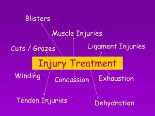
EXERCISE SCIENCE TREATMENT OF AN INJURY p . 73 - 79. Signs of an injury First aid treatment THE SHOULDER JOINT THE KNEE JOINT THE ANKLE JOINT. TREATMENT OF AN INJURY . Signs of an injury: SHARP S welling H eat A ltered appearance and function R ed in colour P ainful.

E N D
EXERCISE SCIENCETREATMENT OF AN INJURYp. 73 - 79 Signs of an injury First aid treatment THE SHOULDER JOINT THE KNEE JOINT THE ANKLE JOINT
TREATMENT OF AN INJURY Signs of an injury: SHARP • Swelling • Heat • Altered appearance and function • Red in colour • Painful
TREATMENT OF AN INJURY First aid treatment immediately following an injury: PIER principle • Pressure – applied with ice; left on 10 – 20 min, similar break time; repeat • Ice – avoid heat during initial days of an injury (will promote swelling) • Elevation during icing to help reduce swelling • Restricted (rested) using tensors and slings
THE SHOULDER JOINT Glenohumeral joint – classified as synovial ball-and-socket joint • the instability of this joint is what permits its excellent mobility • the joint is held together by several ligaments and the tendon of the bicepsbrachiiwhich helps to support the joint anteriorly • very susceptible to injuries from overuse and from heavy physical contact
THE SHOULDER JOINT Biceps Tendinitis • overuse injury from overworking or overloading the joint (not enough rest given) • symptoms: pain on proximal end of the biceps (pain during shoulder or elbow flexion)
THE SHOULDER JOINT Shoulder Separation • occurs at the acromioclavicular joint (tearing of the acromioclavicular ligament) • x-rays are used to determine the severity of the tear • injury results from contact with another player or fall on the shoulder • 3rddegree tears may require surgery; recovery accelerated with physio
THE SHOULDER JOINT Shoulder Dislocation • results when the head of the humerus pops out of the glenoidfossa • this injury results from a hit or fall (tears to the glenohumeralligament and joint capsule) • attempts to relocate the shoulder may results in permanent damage to numerous vital nerves and blood vessels • should only be attempted by qualified personnel • surgery may be required for third-degree dislocations
THE SHOULDER JOINT Rotator Cuff Tears • 4 rotator cuff muscles: supraspinatus, infraspinatus, teres minor, and subscapularis • tears may occur to one or all four of the muscles; 3 of the muscles share a common tendon attachment • causes difficulty and pain when abducting and laterally or medially rotating the shoulder • apply PIER principle to speed up diagnosis and healing
THE KNEE JOINT • This joint is the articulation of the tibia and femur (not fibula) • originally classified as synovial (modified hinge), but now classified as a modified ellipsoid joint because it is now known to slightly rotate medially and laterally
THE KNEE JOINT Knee Ligament Tears • the most common tears result from blows to the lateral side of the knee, which results in damage to the medial side • the first tissue to tear is the joint capsule, and if severe enough, will damage the medial collateral ligament, medial meniscus, and anterior cruciateligament, as well
THE KNEE JOINT Knee Ligament Tears • women are more susceptible to ACL tears and other knee injuries because of their wider Q-angle (quadriceps angle) • the Q-angle is formed in the frontal plane; a line is drawn from: • the centre of the patella to the anterior superior iliac spine • the other is from the tibialtuberosity to the centre of the patella extending up the thigh
THE KNEE JOINT Knee Ligament Tears • the width of the pelvis determines the size of the Q angle • the greater angle in women, causes forces to the concentrated on the ligament each time the knee twists • proper stretching and strengthening will decrease the chance of injury
THE KNEE JOINT OSGOOD-SCHLATTER Syndrome What is it? What causes it? • a result of osteochondritis (a disease of the ossification centres in the bones of young children) • growing pains for the child • in growing child, the growth plates of the tibialtuberosities can become irritated or inflamed if overloaded or overused
THE KNEE JOINT OSGOOD-SCHLATTER SYNDROME What tissues are affected? Who does it affect? • more prevalent in males • running & jumping stresses the patellar tendon and ligament, causing inflammation of the cartilage layer in that growth plate
THE KNEE JOINT OSGOOD-SCHLATTER SYNDROME Future implications & treatment • does not affect growth of child or damage epiphyseal plate • must be diagnosed by physician • follow PIER
THE KNEE JOINT PATELLOFEMORAL SYNDROME (PFS) What is it? What causes it? • gradual onset of anterior knee pain or pain around the patella • the pain is a result of increased or misdirected forces between the patella and femur • aggravated by sports (running, VB, BB...)
THE KNEE JOINT PATELLOFEMORAL SYNDROME (PFS) What tissues are affected? Who does it affect? • usually affects adolescents or young adults, more often women • debate/lack of concensus on factors • overuse, overloading, and misuse of patellofemoral jt. are agreed (researchers)
THE KNEE JOINT PATELLOFEMORAL SYNDROME (PFS) Future implications & treatment • treat with PIER • if pain persists, seek medical care
THE ANKLE JOINT THE ANKLE JOINT • classified as modified hinge • comprised of tibia, fibula and talus
THE ANKLE JOINT Inversion sprains • inversion sprains are common injuries (rolling over on ankle) • ankle is weakest when plantar flexed; thus when you jump and land hard to change direction, the ankle plantar flexes with great force • this injury can affect one or all of the lateral ligaments of ankle • the severity of the sprain dictates the amount of time needed for healing • surgery is rare (even in 3rd degree) • sprains described as low or high; high involve damage to one or both anterior and posterior tibiofibular ligaments • apply PIER
THE ANKLE JOINT Eversionsprains • rare; a very strong deltoid ligament attaches the medial malleolus to 3 bones of the foot, causing tip of medial malleolus to tear off • Pott’sFracture – most severe eversion injury – the tip of the medial malleolus is broken, as is the fibula • Cause: a force on the medial side of ankle, causing deltoid ligament to rip off the tip of the medial malleolus and a break of the fibula • Treatment: case 8 to 12 weeks, then intense physio • Future implications: not career-ending; won’t break in same spot again; care and rehabilitation should be adhered to in order to prevent future weakness