Download
1 / 18
180 likes | 297 Views
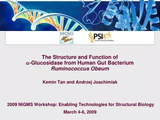
The Structure and Function of -Glucosidase from Human Gut Bacterium Ruminococcus Obeum. Kemin Tan and Andrzej Joachimiak 2009 NIGMS Workshop: Enabling Technologies for Structural Biology March 4-6, 2009. Gut Microbiota. Division. Actinobacteria. Bacterioidetes. Genus. Species.

E N D
The Structure and Function of -Glucosidase from Human Gut Bacterium Ruminococcus Obeum Kemin Tan and Andrzej Joachimiak 2009 NIGMS Workshop: Enabling Technologies for Structural Biology March 4-6, 2009
Gut Microbiota Division Actinobacteria Bacterioidetes Genus Species Clostridium { Strain Firmicutes Eubacterium { Gnavus obeum … etc. Ruminococcus … etc. ATCC 29174 Proteobacteria … etc.
Known Functions • Maturation • Development of innate immunity • Production of essential vitamins etc. Nondigestible food components serve as sources of energy and carbon for the human gut bacteria. The Journal of Nutrition. 2007
Over-Represented Genes Statistics for Some Genomes by COG Catagories Genome NameCOG genes MetabAA Percent MetabCarb Percent MetabLipid Percent Bordetella parapertussis 12822 3654 490 13.41% 197 5.39% 239 6.54% Corynebacterium diphtheriae 1576 174 11.04% 107 6.79% 57 3.62% Cytophaga hutchinsonii 2226 163 7.32% 126 5.66% 97 4.36% ATCC 33406 Enterococcus faecalis V583 2210 180 8.14% 262 11.86% 61 2.76% Escherichia coli K12 3566 367 10.29% 377 10.57% 103 2.89% Geobacter sulfurreducens PCA 2527 190 7.52% 99 3.92% 64 2.53% Haloarcula marismortui 2642 268 10.14% 140 5.30% 95 3.60% ATCC 43049 Listeria innocua 2391 212 8.87% 278 11.63% 59 2.47% Methanocaldococcus jannaschil DSN 2661 1427 106 7.43% 51 3.57% 14 0.98% Porphyromonas gingivalis W83 1233 78 6.38% 59 4.82% 40 3.27% Pseudomonas syringae pv.tomato 4177 458 10.96% 261 6.25% 180 4.31% str.DC3000 Ruminococcus obeum 2393 224 9.36% 245 10.24% 61 2.55% ATCC 29174 Silicibacter pomeroyi DSS 3399 5561 6.36% 204 6.00% 194 5.71% Sulfolobus solfataricus P2 2105 202 9.60% 128 6.08% 86 4.09% Thermoplasma volcanium GSS1 1214 117 9.64% 89 7.33% 50 4.12% Vibrio parahaemolyticus 3259 346 9.80% 207 5.87% 123 3.49% RIMD 2210633
Glycosyl Hydrolases In Ruminococcus obeum ATCC 29174, 245 genes in carbohydrate transport and metabolism, 22 genes as glycosyl hydrolases (GH). GH1, 1 GH2, 2 GH3, 3 GH18, 1 GH20, 1 GH31, 1 (a-glucosidase) GH32, 4 GH42, 3 GH43, 4 GH77, 4
Data collection Phasing Refinement Crystal Structure Determination • X-ray Diffraction Data Collection and Processing: SBCcollect APS, Structural Biology Center, 19ID beamline. HKL3000 program suite data integration and scaling. • Structure Determinaion: HKL3000 program suite 50 out of 54 Se sites located and used in phasing. 46 sites Se sites matched NCS and used for averaging and phase improvement • Model Building: HKL3000 program suite 3 cycles of Arp/warp model building: 1244 out 1332 residues built (93.4%). sequence docked: 1211 residues.
Dimer Structure in Crystal and Solution Calculated monomer molecular weight: 77.4kD, including vector derived residues.
Homologous Structures • Human intestinal maltase-glucoamylase PDB: 2QLY Overall sequence identity: 28% • Sulfolobus solfataricus a-Glucosidase PDB:2G3M Overall sequence identity: 26%
amylose amylopectin Maltase-Glucoamylase(MGAM) (1-4) high activity Sucrase-Isomaltase(SI) (1-4) (1-6) (1-4) (exohydrolases) Human Intestinal MGAM and SI a-Amylase (endohydrolase) Glucose
Catalytic Site Catalytic domain R.obeum a-glucosidase: 366 a.a. Human NtMGAM: 362 a.a. Structural alignment: • 310 a.a. aligned • RMSD: 1.68Å • Sequence identity: 29.6% Catalytic nucleophile: the residue D307 in magenta. Acid/base catalyst (possible): the residue D420 in green.
Substrate Hydrolyzed (mM) Substrate Specificity Maltose Sucrose Lactose • At least a maltase
Access to Catalytic Site • Glucoamylase ?
A Common Enzyme in Gut Microbiota • Iden. Posi. Gap • Coprococcuseutactus ATCC27759 67% 80% 0% • Clostridium sp. L2-50 69% 80% 0% • Clostridium phytofermentans ISDG 59% 75% 0% • Faecalibacteriumprausnitzii M21/2 56% 72% 1% • Clostridium botulinumc str. Eklund 53% 71% 2% • Clostridium perfringens CPE str. F4969 54% 70% 1% • ……….. • Petrotogomobilis SJ95 46% 65% 2%
amylose amylopectin Maltase-Glucoamylase(MGAM) (1-4) high activity Sucrase-Isomaltase(SI) (1-4) (1-6) (1-4) (exohydrolases) Glycosyl Hydrolases glucose
Conclusions 1. PDB: 3FFJ 2. Member of gut microbiota can also utilize digestible carbohydrates. 3. Potential competition between gut micobiota and human host in utilization of carbohydrate resources. 4. Regulation ? ……
Acknowledgements • ANL/MCSG • A. Jochimiak • H. An, • G. Babnigg, • L. Bigelow, • A. Binkowski, • C-s. Chang, • S. Clancy, • G. Cobb, • M. Cuff, • M. Donnelly, • C. Giometti, • W. Eschenfeldt, • Y. Fan, • C. Hatzos, • R. Hendricks • G. Joachimiak, • H. Li, • L. Keigher, • Y-c. Kim, • N. Maltseva, • E. Marland, • S. Moy, • R. Mulligan, • B. Nocek, J. Osipiuk, , M. Schiffer, • A. Sather • G. Shackelford, • L. Stols, • C. Tesar, • R-y. Wu, • L. Volkart, • R-g. Zhang, • M. Zhou, • ANL/SBC • N. Duke, • S. Ginell, • F. Rotella • R.Wilton • Univ. College • London @ EBI, • J. Thornton, • C. Orengo, • M. Bashton, • R. Laskowski, • D. Lee, • R. Marsden, • D. McKenzie, • A. Todd, • J. Watson • Northwestern Univ. • W. Anderson, • O. Kiryukhina • D. Miller, • G. Minasov, • L. Shuvalova, • X. Yang, • Y. Tang • Univ. of Toronto • A. Edwards, • C. Arrowsmith, • A. Savchenko, • E. Evdokimova, • J. Guthrie, • A. Khachatryan, • M. Kudrytska, • T. Skarina, • X. (Linda) Xu • Washington • Univ. • D. Fremont, • T. Brett, • C. Nelson, • Univ. of Chicago • O. Schneewind, • D. Missiakas, • P. Gornicki, • S. Koide, ITCSG • W-j. Tang, • B. Roux, • J. L. Robertson • M.R. Rosner, • T. Kossiakoff, ITCSG • V. Tereshko, • G. Montelione, • Ruthgers Univ. NESGC • T. Terwilliger, • Los Alamos, ITCSG • Z. Derewenda, Univ. • of Virginia, ITCSG • Z. Dauter, NCI • J. Liang, Univ. • of Illinois • D. Sherman, U. Michigan • Univ. of Texas • SWMC • Z. Otwinowski, • D. Borek, • A. Kudlicki, • A. Q. Mei, • M. Rowicka • Univ. of Virginia • W. Minor, • M. Chruszcz, • M. Cyborowski, • M. Grabowski, • P. Lasota, • P. Miles, • M. Zimmerman, • H. Zheng • Funding: NIH and DOE • 17 • 17