Download
1 / 19
190 likes | 211 Views
Explore the methodology of generating realistic synthetic medical images and associated segmentations for object evaluation. Techniques include probability distribution sampling, deformation of template geometry, and image processing. Developed by James Z. Chen and team from the Medical Image Display & Analysis Group at University of North Carolina.

E N D
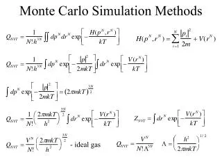
Medical Image Synthesis via Monte Carlo Simulation An Application of Statistics in Geometry & Building a Geometric Model with Correspondence James Z. Chen, Stephen M. Pizer, Edward L. Chaney, Sarang Joshi, Joshua Stough Presented by: Joshua Stough Medical Image Display & Analysis Group, UNC midag.cs.unc.edu
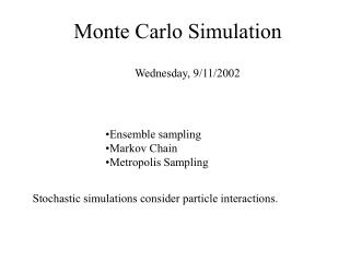
Population Simulation Requires Statistical Profiling of Shape Goal:Develop a methodology for generating realistic synthetic medical images AND the attendant “ground truth” segmentations for objects of interest. Why:Segmentation method evaluation. How: Build and sample probability distribution of shape.
Basic Idea • New images via deformation of template geometry and image. • Characteristics • Legal images represent statistical variation of shape over a training set. • Image quality as in a clinical setting. Ht
The Process James Chen
Registration • Registration – Composition of Two Transformations • Linear – MIRIT, Frederik Maes • Affine transformation, 12 dof • Non-linear–Deformation Diffeomorphism, Joshi • For all It , It Ht(I0) and St Ht(S0)
Consequence of an Erroneous Ht James Chen
Generating the Statistics of Ht James Chen
Fiducial Point Model • Htis locally correlated • Fiducial point choice via greedy iterative algorithm • Ht' determined by Joshi Landmark Deformation Diffeomorphism • The Idea: Decrease
FPM Generation Algorithm • Initialize {Fm} with a few geometrically salient points on S0; • Apply the training warp function Ht on {Fm} to get the warped fiducial points: Fm,t = Ht(Fm); • Reconstruct the diffeomorphic warp field H't for the entire image volume based on the displacements {Fm,t – Fm}; • For each training case t, locate the point pt on the surface of S0 that yields the largest discrepancy between Ht and H't; • Find most discrepant point p over the point set {pt} established from all training cases. Add p to the fiducial point set; • Return to step 2 until a pre-defined optimization criterion has been reached.
A locally accurate warp via FPM landmarks Volume overlap optimization criterion tracks mean warp discrepancy Under 100 fiducial points, of thousands on surface ATLAS WARP TRAINING
Human Kidney Example • 36 clinical CT images in the training set • Monotonic Optimization • 88 fiducial points sufficiently mimick inter-human rater results (94% volume overlap)
Fiducial Point Model Is an Object Representation with Positional Correspondence • Positional correspondence is via the H' interpolated from the displacements at the fiducial points • The correspondence makes this representation suitable for statistical analysis
Statistical Analysis of the Geometry Representation James Chen
Principal Components Analysis of the FPM Displacements • Points in 3M-d space • Analyze deviation from mean • Example: first seven modes of FPM cover 88% of the total variation.
-2 +1 -1 +2 ATLAS I II III MEAN Modes of Variation – Human Kidney
Results James Chen
Miscellaneous • National Cancer Institute Grant P01 CA47982 References Gerig, G., M. Jomier, M. Chakos (2001). “Valmet: A new validation tool for assessing and improving 3D object segmentation.” Proc. MICCAI 2001, Springer LNCS 2208: 516-523. Cootes, T. F., A. Hill, C.J. Taylor, J. Haslam (1994). “The Use of Active Shape Models for Locating Structures in Medical Images.” Image and Vision Computing12(6): 355-366. Rueckert, D., A.F. Frangi, and J.A. Schnabel (2001). “Automatic Construction of 3D Statistical Deformation Models Using Non-rigid Registration.” MICCAI 2001, Springer LNCS 2208: 77-84. Christensen, G. E., S.C. Joshi and M.I. Miller (1997). “Volumetric Transformation of Brain Anatomy.” IEEE Transactions on Medical Imaging16: 864-877. Joshi, S., M.I. Miller (2000). “Landmark Matching Via Large Deformation Diffeomorphisms.” IEEETransactions on Image Processing. Maes, F., A. Collignon, D. Vandermeulen, G. Marchal, P. Suetens (1997). “Multi-Modality Image Registration by Maximization of Mutual Information.” IEEE-TMI16: 187-198. Pizer, S.M., J.Z. Chen, T. Fletcher, Y. Fridman, D.S. Fritsch, G. Gash, J. Glotzer, S. Joshi, A. Thall, G. Tracton, P. Yushkevich, and E. Chaney (2001). “Deformable M-Reps for 3D Medical Image Segmentation.” IJCV, submitted.