Download
1 / 10
100 likes | 112 Views
This article explores various methods for diagnosing pneumonia, including blood and phlegm samples, C-reactive protein expression, and chest radiographs. Future research on improving radiograph quality is also discussed.

E N D
Methods in diagnosing pneumonia for the improvement of radiographs Daisy Ochoa University of California, Merced
Background Fig 1. Consolidation: Inflammation of the lung tissue [4]. • Community acquired Pneumonia (CAP) is a respiratory infection of the lungs in which the air sacks become swollen due to foreign micro-organisms: bacteria and viruses [1]. • One of the leading causes of death with an estimation of about 12 cases per 1000 adults each year [3]. • 600 people under the age of 65 were admitted to the hospital for CAP [5] • Chest x-rays, blood and phlegm samples are used in the diagnosis
Methods • Process of Identifying the microorganism: Blood and phlegm samples are collected for lab tests. White blood cell count (WBC), C-reactive protein (CRP), and the rate of red blood cell’s sedimentation is also done in the lab [3]. • ELISA test
Results from the ELISA test • Blood samples: 3 patients out of the 10 test positive for pneumococcal pneumonia. The blood cultures drops that were used in the ELISA did show pneumococcal capsular antigens. • Other samples, such as sputum, showed that the infections were caused by Staphylococcus aureus in patients with flu-like symptoms.
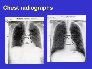
C-reactive Protein • High expression of CPR shown by the ELISA test. A correlating factor that is used as a diagnosing tool for pneumonia. • CRP is a protein released as a inflammation signaling response from the liver. • Consolidation of the lungs results in high expression of CRP [3]. Fig 3. CRP structure Fig 4. Patchy consolidation CXR [4]
More Methods . . . • Radiographs from chest x-ray (CXR) or a computed tomography scan (CT scan) are exposed to patients with suspected pneumonia. • Upright position with protective radiation shielding • Radiographs: short x-ray pulse is exposed to the patient in which the bones absorb due to the calcium's high absorbency [2]. Bone and air are seen clearly on radiographs unlike tissue due to the density differences of each tissue. • Radiographs sometimes are unclear and difficult to read due to the interference of consolidation.
Future research • Silicon based detectors (lower radiation exposure) vs silver based detectors • Researchers Hilt, B., Fessler, P., & Prevot, G conducted single x-ray exposures to phantom body parts using regular silver based radiation and silicon based detector of x-rays but produce a low quality image. • A future goal: use lower exposure of radiation but to produce high quality images. • Future holds improvement to the lab apparatusfor researchers to increasing the spatial resolution, remove unwanted regions, and keep a good contrast within the images.
In summary • Multiple methods used in the diagnosis of pneumonia • Blood samples • Phlegm samples • CRP expression • ELISA test • Chest x-rays and CT scans • Future research for radiograph quality
References • Hilt, B., Fessler, P., & Prevot, G. (2000). New quantum detection system for very low dose x-ray radiology. Nuclear Instruments and Methods in Physics Research, A(421), 38-44. • Hayden, G. E., & Wrenn, K. W. (2009). Chest radiograph vs. computed tomography scan in the evaluation for pneumonia. The Journal of Emergency Medicine, 36(3), 266-270. doi: 10.1016/j.jemermed.2007.11.042 • Castro-Guardiola, A., Armengou-Arxe ́et al. (2000). Differential diagnosis between community-acquired pneumonia and non- pneumonia diseases of the chest in the emergency ward. European Journal of Internal Medicine, 11, 334-339. • Das, D., & Howlett, D. C. (2009). Chest x-ray manifestations of pneumonia. 27(10), 453-455. • Tang, C. M., & Macfarlane, J. T. (1993). Early management of younger adults dying of community acquired pneumonia. Respiratory Medicine, 87, 289-294.