Download
1 / 34
350 likes | 527 Views
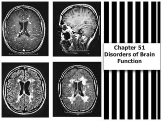
Chapter 36 Disorders of Brain Function. Hypoxia and Ischemia. Hypoxia causes ATP depletion or “power failure” Aerobic metabolism stops less ATP is produced Na + /K + ATPase cannot run fast enough Cell swells up with water Anaerobic metabolism used lactic acid produced

E N D
Hypoxia and Ischemia • Hypoxia causes ATP depletion or “power failure” • Aerobic metabolism stops less ATP is produced • Na+/K+ ATPase cannot run fast enough • Cell swells up with water • Anaerobic metabolism used lactic acid produced • Acid damages cell membranes, intracellular structures, and DNA
Hypoxia and Ischemia (cont.) Ischemia also interferes with: • Delivery of energy stores (e.g., glucose) • Damage to blood vessels • Vasomotor paralysis • Vasoconstriction • Changes in blood • Desaturation • Clotting • Sludging
Question What two substances are needed by the mitochondria in order to produce ATP? • O2 and CO2 • Glucose and O2 • Glucose and fatty acids • Proteins and monosaccharides
Answer • Glucose and O2 Rationale:Glucose and oxygen are necessary for ATP production. Without ATP, no physiologic work can be done—the cells, and eventually the organism, will die. When oxygen is not available, anaerobic pathways are used, creating lactic acid that also damages the cell.
Calcium Cascade • Ischemia depolarization • Depolarization glutamate release • Glutamate calcium cascade • Calcium influx depolarization
Intracranial Pressure (ICP) • Compartment syndrome in the skull • Intracranial pressure greater than arterial blood pressure • Arteries collapse; blood flow to brain cut off • Brain swelling • Vasogenic: extracellular fluid • Cytotoxic: intracellular fluid • Hydrocephalus: cerebrospinal fluid • Tumors
Brain Herniation • Increased intracranial pressure pushes the brain out of position • Brain tissue is compressed into the center of the brain (2), against bone (4) or against rigid folds of the dura mater (1, 3) • Compression of the oculomotor nerve is an early sign
Traumatic Brain Injury • Primary injuries—due to impact • Microscopic damage: concussion, diffuse axonal injury • Contusions • Secondary injuries—due to: • Hemorrhage • Ischemia • Infection • Increased intracranial pressure
Question Tell whether the following statement is true or false. Increased ICP results in primary brain injury.
Answer False Rationale:Increased pressure in the brain leads to secondary brain injury (there’s nowhere for the pressure to be released because the brain is encased in bone). Primary brain injury is caused by trauma.
Hematoma—Broken Blood Vessels • Epidural space: meningeal arteries • Rapid bleeding; unconsciousness may be followed by brief lucid period • Dura mater • Subdural space: bridging veins • Slower bleeding; gradual development over days or weeks
Cerebral Blood Flow Middle cerebral artery • Internal carotid arteries branch into: • Anterior cerebral arteries • Medial and superior surfaces of brain; frontal lobes • Middle cerebral arteries • Lateral surfaces of brain: face and arm motor and sensory cortexes, optic radiations, speech centers Anterior cerebral artery Brain (lateral view) Internal carotid artery
Cerebral Blood Flow (cont.) • The basilar artery runs up to the back of the brain • It splits to form the two posterior cerebral arteries • They supply the medulla, pons, cerebellum, midbrain, occipital lobes, temporal lobes, thalamus Posterior cerebral artery Basilar artery
Brain: ventral view Circle of Willis • Anterior communicating artery • Connects right and left anterior cerebral arteries • Blood from one carotid can cross over to supply the other side of the brain • Posterior communicating arteries • Connect the posterior and middle cerebral arteries • Blood from the basilar artery can run forward and supply the front of the brain
Question Which of the following blood vessels ensures collateral circulation in the brain? • Internal carotid arteries • Cerebral arteries • Basilar arteries • Circle of Willis
Answer • Circle of Willis Rationale:The circle of Willis connects the right and left anterior cerebral arteries and the posterior and middle cerebral arteries. Blood from one carotid can cross over to supply the other side of the brain; blood from the basilar artery can run forward and supply the front of the brain.
Stroke • Stroke = “brain attack” • Ischemic stroke • Large vessel (thrombotic) • Small vessel (lacunar infarct) • Cardiogenic embolic • Hemorrhagic stroke • Transient ischemic attacks (“brain angina”)
Excitotoxicity • Neuron firing releases glutamate • Causes neighboring neurons to fire • Spreading injury across the ischemic area
Discussion Mr. X has cor pulmonale. Mr. Y has a left ventricular aneurysm. Questions: • Which of them is more likely to have a stroke? • Which is more likely to have a pulmonary embolism?
Aneurysmal Subarachnoid Hemorrhage • Aneurysm • Sudden-onset headache with nausea, vomiting, dizziness • Hemorrhage • Sudden severe headache, neck stiffness, photophobia, vision and motor problems • Complications • Rebleeding, vasospasm and ischemia, hydrocephalus, hypothalamus dysfunction, seizures
Brain Tumors • Focal disturbances • Dysfunction of particular brain areas • Seizures, hallucinations, weakness or palsies in specific areas, sensory deficits • Generalized disturbances • Increased intracranial pressure: headache, vomiting, visual problems
Seizures • Spontaneous nerve firing • Provoked seizures • Fever • Electrolyte imbalances (hypocalcemia, alkalosis) • Hypoglycemia • CNS infection or damage • Unprovoked seizures: cause unknown
Epileptic Syndromes • Partial seizures • Begin in one cerebral hemisphere • Secondarily generalized seizures • Begin in one hemisphere and spread to other • Generalized seizures • Involve both hemispheres
Kinds of Seizures • Absence (petit mal): disturbances in consciousness • Atonic: loss of muscle tone • Myoclonic: muscles contract • Tonic-clonic (grand mal): muscle contraction and loss of consciousness • Generalized convulsive status epilepticus: seizures continue without recovery between them
Question Which type of seizure affects only one cerebral hemisphere? • Partial • Secondarily generalized • Generalized • All of the above
Answer • Partial Rationale:Partial seizures affect one cerebral hemisphere; secondarily generalized seizures begin in one hemisphere and then spread to the other side; generalized seizures involve both hemispheres.
Dementias • Many dementias are associated with abnormal inclusions in the brain • Alzheimer disease: amyloid plaques • Pick disease: Pick bodies • Prion diseases: prion proteins • Creutzfeldt-Jakob disease
Alzheimer Disease • Amyloid-beta protein-forming plaques • Neurofibrillary tangles • Decreased acetylcholine production
amyloid Alzheimer Disease precursor protein in normally Alzheimer disease soluble amyloid b protein fragments stick together to form fibrils cleared away amyloid plaques
Stages of Alzheimer Disease • First: short-term memory loss • Second: confusional stage • Disorientation, lack of insight, impaired hygiene and language use, sundown syndrome • Third: incontinence, inability to recognize family and friends
Other Causes of Dementia • Microinfarcts:vascular dementia • Vitamin B12 deficiency: Wernicke-Korsakoff syndrome • Inherited atrophy of brain structure:Huntington disease
Question Which cause of dementia is vascular in nature? • Alzheimer • Microinfarcts • Vitamin B12 deficiency • Inherited
Answer • Microinfarcts Rationale:Small infarctions cause blood flow to be cut off to certain areas of the brain, causing tissue death. Depending on the extent of the infarctions, the dementia may be more or less severe.