Download
1 / 30
310 likes | 727 Views
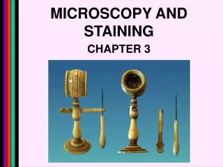
Microbiology and Microscopy Experiment 4 – Cappuccino and Sherman. Professor Sidelsky psidelsky@gmail.com. Introducing your microscope. Always clean lenses before starting Always with both hands supporting base Store with arm facing the door. Cover

E N D
Microbiology and MicroscopyExperiment 4 – Cappuccino and Sherman Professor Sidelsky psidelsky@gmail.com
Introducing your microscope • Always clean lenses before starting • Always with both hands supporting base • Store with arm facing the door. Cover • Focus with scanning and low before proceeding to high and oil • Learn the most effective ways to regulate and manage light
Eyepiece( Ocular) Binocular –two eyepieces – magnification 10x Objective lenses – 4( see chart) Condenser lens – collects and directs light through the lenses Condenser adjustment – raises and lowers the condenser lens for better focus Mechanical stage adjustment – Knobs that move the mechanical stage Coarse adjustment – used with scanning and low power Fine adjustment – used with high and oil Microscope Parts LENSES ADJUSTMENTS
Checklist • Find the following parts on your microscope,and be sure to know the functions of each: Coarse adjustment knob Fine adjustment knob Eyepieces Objective lenses- 4x, 10x, 40x, 100x oil Light source (lamp) On-off knob for light Condenser adjustment knob Diaphragm lever
Olympus web site on the anatomy of a microscope • http://www.olympusmicro.com/primer/anatomy/anatomy.html
Eyepiece • Adjustment of the Interpupillary Distance The interpupillary distance is the distance between the centers of your two pupils. The distance between the two eyepieces of the binocular microscope must correspond to your interpupillary distance. • Each person has his or her own interpupillary distance and the microscope must be adjusted for your specific distance. • This is true of any binocular microscope. During your first session with a new microscope you must determine the correct interpupillary distance and set the microscope for that distance. If you look through the eyepieces and see two images, the interpupillary distance is not correct. To correct it, slide the eyepieces closer together or farther apart until the two fields merge to form a single circle of light. The interpupillary distance is now correct for you.
Mechanical Stage and Adjustments • Holds slide in position • Movement of slide on X and Y axis with adjustment knobs • Permits student to locate specific site on slide
Condenser lens and iris diaphragm Condenser lens Iris diaphragm
Coarse and fine adjustment • Use the coarse adjustment to assist you in focusing on scanning and low – Do not use the coarse adjustment on high and oil
Microscope terms • Microscope terms • Magnification • Total magnification • Resolution • Depth of field • Size of field • Working distance • Parfocal • Parcentric
Total Magnification • The magnification of the objective lens X the magnification of the ocular( 10)
Resolution • Resolution is the ability to see two points as separate entities • Resolution is determined by the magnification and the numerical aperture – The numerical aperture is the opening at the bottom of the lens
Numerical aperture • A measure of the lenses ability to gather light and focus an image at a precise focal lengthh
Terms • Size of field – the diameter of the field of view at different magnifications • Depth of field – the depth of the image through which it is possible to focus • Working distance - the distance between the stage and the bottom of the lens • Ease of focus – ability to bring image into focus
More Terms • Parfocal – The quality of a microscope that enables one change from a lower power to a higher power and still retain the focus • Parcentric – If the image is basically in the center of the field of view – it will remain in the center even if the magnification is changed.
Practice Slide for technique • Use a prepared slide to view bacterial cells. Place it on the Stage • Observe the slide on scanning, low, and high power. • Adjust the light and draw on high power.
High Power • Always begin focus on scanning or low. Look over the slide to choose and optimal section for viewing. • If your focus is sharp and clear, turn the revolving nosepiece to high (blue) • Use the fine adjustment, focus carefully – you are very close to the slide.
Light Adjustment( Three ways to adjust the light) • Iris Diaphragm – lever under the stage. This works like the iris of the eyes • Condenser lens – collects and directs the light. Use the condenser adjustment to move the lens closer to and further from the stage. • Light intensity adjustment( diopter) can also be checked.
Oil immersion lens • This lens provides the best resolution and the highest magnification ( 1000x) • Focus first on high power. Tweak your focus until it is perfect with the fine adjustment. • Turn the revolving nosepiece until it is between the high and oil immersion lens, use oil dropper to place a drop over the center of the slide. Slowly turn lens into the oil. Then again use the fine adjustment to make the image clear.
Pond Water • Practice using microscope with the slides of pond water. • Draw your slides on scanning, low, high, and oil • Review your procedures for using coarse and fine adjustment, the condenser knobs, and mechanical stage
Cleaning Up 1. Put low power objective lens in place. 2. Lower stage all the way down. 3. Clean oil off immersion lens. 4. Remove slide and clean off oil. 5. Turn light off. 6. Unplug and cover.