Collapsing Trachea
Collapsing Trachea. Mark Bohling, DVM Diplomate, American College of Veterinary Surgeons Assistant Professor of Surgery University of Tennessee College of Veterinary Medicine. What is Tracheal Collapse?. Normal airflow dynamics in respiration Inspiration

Collapsing Trachea
E N D
Presentation Transcript
Collapsing Trachea Mark Bohling, DVM Diplomate, American College of Veterinary Surgeons Assistant Professor of SurgeryUniversity of TennesseeCollege of Veterinary Medicine
What is Tracheal Collapse? • Normal airflow dynamics in respiration • Inspiration • Expansion of chest by muscles of respiration • Pressure gradient - chest negative • Effect on the air conduit: • Thorax - expansion • Neck - compression • Expiration • Reverse effects
History of Collapsing Trachea in Veterinary Medicine • Described as early as 1960 • Review of early treatments • Single plastic tube • Ventral chondrotomy • Modified ventral chondrotomy • Dorsal membrane plication
Tracheal Collapse in Other Species • Tracheal collapse in human beings • History • Dates to 1930’s • Similarities • Softening of tracheal cartilage • Lateral collapse (same as dorsoventral in dogs) • Differences • Classification • Primary vs secondary collapse • Pediatric vs adult collapse
Tracheal Collapse in Other Species • Tracheal collapse in large animals • Horses • Congenital • Secondary to laryngeal paralysis • Cattle • Acquired neonatal • Tracheal collapse in birds • Bordetella avium in turkeys
Tracheal Collapse in the Dog • Miniature breeds • Middle aged to older • Other risk factors • More pronounced in obese individuals
Levels of Collapse Normal G1 G2 G3 G4
Clinical Signs • Chronic, dry nonproductive cough (honking) • Intermittent dyspnea (worsens with excitement) • Cyanosis & syncope in severe cases • Inspiratory/ expiratory dyspnea • Prone to heat stroke
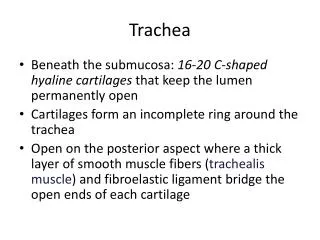
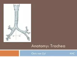
Pathophysiology • Disease causes the trachea rings to weaken • Dorsal ligament and trachealis muscle weaken and stretch • Trachea changes from oval tube to a flattened conduit
Etiology • Congenital • Nutritional tracheomalacia • Obesity • Bacterial infection • Neurologic • Chronic airway disease • Idiopathic – “who knows why”
Diagnosis • Tracheal palpation • Radiographs (inspiratory / expiratory ) • Fluoroscopy • Tracheoscopy
Medical Management • Cough suppression(Hydrocodone, butorphanol) • Bronchial dilators(Aminophylline, terbutaline) • Sedation(Acepromazine) • Weight loss
Medical Management • Help control symptoms • Can not be cured • Disease usually progressive
Surgical Correction • External stenting with plastic rings
Before After
Surgical Correction • External spiral stent
External Stent Complications • Collapse between rings
External Stent Complications • Damage to recurrent laryngeal nerve
External Stent Complications • Interruption of tracheal bloodsupply
Internal Stenting • What is a stent? • History of stenting • History of tracheal stenting • Modern stents and stent materials • Stents in veterinary medicine
Ultraflex® Stent • Radiopaque, self-deployed • 4 - 8 cm length, 10 - 20mm diameter • Made of nitinol (nickel-titanium alloy) • Proximal or distal deployment • Single strand, open loop knitted design (flexible, contourable) Boston Scientific/ Microvasive.
SmartStent® • Nitinol tube • Laser cut • No overlapping wires • Less breakage in human vascular applications • Cordis Endovascular®
Infiniti Stent • Also nitinol • Single woven wire • Only stent produced exclusively for vet use • Claims as yet unproven
Stent Placement • Stent deployed under fluoroscopic guidance • Target – 5mm cranial to bifurcation • Placement checked with tracheoscopy
Postoperative Care • Perioperative antibiotics • Corticosteroids for 7 days • Sedation • Cough suppression • 24 hours oxygen if needed • Humidification
Stent Results • The little girl with the curl syndrome… • Good outcomes… • Immediate improvement • Breathing near normal • Mild chronic cough • And the not-so-good outcomes…
Stent Complications • Stent fracture • Granulation in stent • Tracheal exudate • Additional collapse at ends of stent
Stent Fracture • Originally thought to be due to bending stresses • All brands/types of nitinol stents can fracture - there is NO unbreakable stent • At this time, removal is best option - BUT - not for the fainthearted!