Download
1 / 49
500 likes | 584 Views
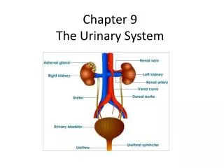
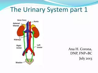
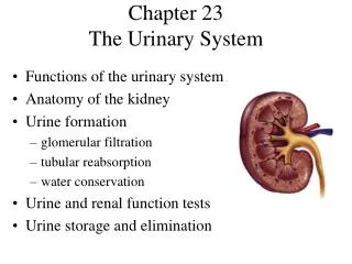
The Urinary System Chapter 15 Part B. Ureters. Slender tubes attaching the kidney to the bladder Continuous with the renal pelvis Enter the posterior aspect of the bladder Runs behind the peritoneum Peristalsis aids gravity in urine transport. Organs of the Urinary System. Figure 15.1a.

E N D
Ureters • Slender tubes attaching the kidney to the bladder • Continuous with the renal pelvis • Enter the posterior aspect of the bladder • Runs behind the peritoneum • Peristalsis aids gravity in urine transport
Organs of the Urinary System Figure 15.1a
Urinary Bladder • Smooth, collapsible, muscular sac • Temporarily stores urine • Trigone—triangular region of the bladder base • Three openings • Two from the ureters • One to the urethra • In males, the prostate gland surrounds the neck of the bladder • Retroperitoneal – posterior to pubic symphysis Figure 15.6
Female Urinary Bladder and Urethra Figure 15.6
Urinary Bladder Wall • Three layers of smooth muscle collectively called the detrusor muscle • Mucosa made of transitional epithelium • Walls are thick and folded in an empty bladder • Bladder can expand significantly without increasing internal pressure
Urinary Bladder Capacity • A moderately full bladder is about 5 inches long and holds about 500 mLof urine • Capable of holding twice that amount of urine
Distended v. Empty Bladder in an Adult Man Note the expansion ability!! Figure 15.7
Renal Calculi aka “Kidney Stones” • May be found throughout the Urinary System • Remove through surgery or lithotripsy (ultrasound) • Form due to concentrated urine • Bacterial infections • Diet – alkaline urine; vegetarian diet
Kidney stones • Video Clip: Symptoms & Treatment of Kidney Stones
Urethra • Thin-walled tube that carries urine from the bladder to the outside of the body by peristalsis • Release of urine is controlled by twosphincters: • Internal urethral sphincter • Involuntary and made of smooth muscle • External urethral sphincter • Voluntary and made of skeletal muscle
Female Urinary Bladder and Urethra Figure 15.6
Urethra Gender Differences • Length • Females – 3–4 cm ( approx. 1 inch) • Males – 20 cm (approx. 8 inches) • Location • Females – anterior to and along wall of the vagina • Males – through the prostate and penis • 3 regions: prostatic, membranous, spongy (penile)
Urethra Gender Differences • Function • Females – only carries urine • Males – carries urine and is a passageway for sperm cells • Urethritis – inflammation of urethra • Cystitis – inflammation of bladder • Pyelonephritis – inflammation of kidney
Micturition (Voiding) • Both sphincter muscles must open to allow voiding • The internal urethral sphincter is relaxed after stretching of the bladder • Activation is from an impulse sent to the spinal cord and then back via the pelvic splanchnicnerves • Urine is forced past the internal urethra sphincter and the person feels the urge to void • The external urethral sphincter must be voluntarily relaxed
Micturition (Voiding) • Incontinence • <2 years old • Pressure (pregnancy) • Nervous system problem • Retention • After surgery (anesthesia) • Hyperplasia (male prostate)
Fluid, Electrolyte, and Acid-Base Balance • Blood composition depends on three factors • Diet • Cellular metabolism • Urine output
Fluid, Electrolyte, and Acid-Base Balance • Kidneys have four roles in maintaining blood composition • Excretion of nitrogen-containing wastes (previously discussed) • Maintaining water balance of the blood • Maintaining electrolyte balance of the blood • Ensuring proper blood pH
Maintaining Water Balance • Normal amount of water in the human body • Young adult females – 50% • More fat, less muscle • Young adult males – 60% • Babies – 75% • Old age – 45% • Water is necessary for many body functions and levels must be maintained
Distribution of Body Fluid • ICF - Intracellular fluid • Fluid inside cells • About 2/3 of body fluid • ECF - Extra cellular fluid • Fluid outside cells • 1/3 of body fluid • Interstitial fluid • Blood plasma • CSF • Serous fluid Figure 15.8
Major Fluid Compartments of the Body Figure 15.8
The Continuous Mixing of Body Fluids Figure 15.9
The Link Between Water and Salt • Solutes in the body include electrolytes like sodium, potassium, and calcium ions • Changes in electrolyte balance (Na, K, Ca) causes water to move from one compartment to another • Alters blood volume and blood pressure • Can impair the activity of cells • Muscle, nerves
Maintaining Water Balance • Water intake must equal water output • Sources for water intake • Ingested foods and fluids • Water produced from metabolic processes (10%) • Sources for water output • Vaporization out of the lungs • Lost in perspiration • Leaves the body in the feces • Urine production • Kidneys adjust for water balance • Thirst mechanism is the driving force for water intake
Water Intake and Output Figure 15.10
Maintaining Water Balance • Dilute urine is produced if water intake is excessive • Less urine (concentrated) is produced if large amounts of water are lost—dehydration!! • Proper concentrations of various electrolytes must be present • Controlled by kidneys
Regulation of Water and Electrolyte Reabsorption • Osmoreceptors • Specialized cells in the hypothalamus • React to changes in blood composition by becoming more active • blood volume causes osmoreceptors in hypothalamus to send nerve impulse to pituitary
Regulation of Water and Electrolyte Reabsorption • Regulation occurs primarily by hormones: • Antidiuretic hormone (ADH) (from posterior pituitary) • Prevents excessive water loss in urine • Causes the kidney’s collecting ducts to reabsorb more water • Diabetes insipidus • Occurs when ADH is not released • Leads to huge outputs of dilute urine
Regulation of Water and Electrolyte Reabsorption • Regulation occurs primarily by hormones (continued) • Aldosterone (from adrenal cortex) • Regulates sodium ion content of ECF • Sodium is the electrolyte most responsible for osmotic water flow • Aldosterone promotes reabsorption of sodium ions • Remember, water follows salt!
Regulation of Water and Electrolyte Reabsorption • Renin-angiotensinmechanism: • Mediated by the juxtaglomerular (JG) apparatus of the renal tubules • When cells of the JG apparatus are stimulated by low blood pressure, the enzyme renin is released into blood • Renin produces angiotensinII • Angiotensin causes vasoconstriction and aldosteronerelease • Result is increase in blood volume and blood pressure
Falling systemic bloodpressure volume (+) Reduced filtrate volumeor solute content in renaltubules Inhibits baroreceptorsin blood vessels Hypothalamicosmoreceptors (+) (+) (+) (+) Sympathetic nervoussystem Posterior pituitary JG cells of kidneys Releases (+) ADH (antidiuretichormone) Release Systemic arterioles Causes (+) Renin Collecting ductsof kidneys Vasoconstriction Leads to Results in Causes Peripheral resistance Angiotensin IIformed in blood H2O reabsorption (+) (+) (+) Systemic arterioles Adrenal cortex Causes Secretes Vasoconstriction Aldosterone Results in Targets Peripheral resistance Kidney tubules Causes Na+reabsorption (andH2O absorption) Results in Blood volume KEY: (+) = stimulates Rising blood pressure Renin-angiotension system Neural regulation (sympatheticnervous system effects) Effects of ADH release Maintaining Water and Electrolyte Balance
Falling systemic bloodpressure volume (+) Reduced filtrate volumeor solute content in renaltubules Hypothalamicosmoreceptors Inhibits baroreceptorsin blood vessels (+) (+) (+) (+) Sympathetic nervoussystem Posterior pituitary JG cells of kidneys (+) Release Systemic arterioles Causes Renin Vasoconstriction Leads to Results in Peripheral resistance Angiotensin IIformed in blood (+) KEY: (+) = stimulates Renin-angiotension system Neural regulation (sympatheticnervous system effects) Effects of ADH release Maintaining Water and Electrolyte Balance Figure 15.11, step 5
Falling systemic bloodpressure volume (+) Reduced filtrate volumeor solute content in renaltubules Inhibits baroreceptorsin blood vessels Hypothalamicosmoreceptors (+) (+) (+) (+) Sympathetic nervoussystem Posterior pituitary JG cells of kidneys Releases (+) ADH (antidiuretichormone) Release Systemic arterioles Causes (+) Renin Collecting ductsof kidneys Vasoconstriction Leads to Results in Causes Peripheral resistance Angiotensin IIformed in blood H2O reabsorption (+) (+) (+) Systemic arterioles Adrenal cortex Causes Secretes Vasoconstriction Aldosterone Results in Targets Peripheral resistance Kidney tubules Causes Na+reabsorption (andH2O absorption) KEY: (+) = stimulates Renin-angiotension system Neural regulation (sympatheticnervous system effects) Effects of ADH release Maintaining Water and Electrolyte Balance
Falling systemic bloodpressure volume (+) Reduced filtrate volumeor solute content in renaltubules Inhibits baroreceptorsin blood vessels Hypothalamicosmoreceptors (+) (+) (+) (+) Sympathetic nervoussystem Posterior pituitary JG cells of kidneys Releases (+) ADH (antidiuretichormone) Release Systemic arterioles Causes (+) Renin Collecting ductsof kidneys Vasoconstriction Leads to Results in Causes Peripheral resistance Angiotensin IIformed in blood H2O reabsorption (+) (+) (+) Systemic arterioles Adrenal cortex Causes Secretes Vasoconstriction Aldosterone Results in Targets Peripheral resistance Kidney tubules Causes Na+reabsorption (andH2O absorption) Results in Blood volume KEY: (+) = stimulates Rising blood pressure Renin-angiotension system Neural regulation (sympatheticnervous system effects) Effects of ADH release Maintaining Water and Electrolyte Balance
Maintaining Acid-Base Balance in Blood • Blood pH must remain between 7.35 --7.45 to maintain homeostasis • Alkalosis – pH above 7.45 • Acidosis – pH below 7.35 • Physiological acidosis – pH between 7.35 -- 7.0 • Most ions originate as by-products of cellular metabolism
Maintaining Acid-Base Balance in Blood • Acids produced by the body • Phosphoric acid, lactic acid, fatty acids • Carbon dioxide forms carbonic acid • Base • Ammonia • Most acid-base balance is maintained by the kidneys
Maintaining Acid-Base Balance in Blood • Other acid-base controlling systems: • Blood buffers • Buffer = molecules that prevent dramatic changes in H+ concentration when acids & bases are added • Acids = proton (H+) donors • Bases = proton acceptors • Respiration
Blood Buffers • Acids are proton (H+) donors • Strong acids dissociate completely and liberate all of their H+ in water • Weak acids, such as carbonic acid, dissociate only partially • Bases are proton (H+) acceptors • Strong bases dissociate easily in water and tie up H+ • Weak bases, such as bicarbonate ion and ammonia, are slower to accept H+
Dissociation of Strong and Weak Acids Figure 15.12
Blood Buffers • Molecules react to prevent dramatic changes in hydrogen ion (H+) concentrations: • Bind to H+ when pH drops • Release H+ when pH rises • Three major chemical buffer systems: • Bicarbonate buffer system • Phosphate buffer system • Protein buffer system
The Bicarbonate Buffer System • Mixture of carbonic acid (H2CO3) and sodium bicarbonate (NaHCO3) • Carbonic acid is a weak acid that does not dissociate much in neutral or acid solutions • Bicarbonate ions (HCO3–) react with strong acids to change them to weak acids HCl + NaHCO3 H2CO3 + NaCl strong acid weak base weak acid salt
The Bicarbonate Buffer System • Carbonic acid dissociates in the presence of a strong base to form a weak base and water NaOH + H2CO3 NaHCO3 + H2O strong base weak acid weak base water
The Bicarbonate Buffer System • Biucarbonate ions react with strong acids to create weak acids: HCl + NaHCO3→ H2CO3 + NaCl strong acid weak base weak acid salt • Carbonic acid dissociates in the presence of a string base to form a weak base and water: NaOH + H2CO3→ NaHCO3 + H2O strong base weak acid weak base water
Respiratory System Controls of Acid-Base Balance • Carbon dioxide in the blood is converted to bicarbonate ion and transported in the plasma • Increases in hydrogen ion concentration (acidosis) produces more carbonic acid • Excess hydrogen ion can be “blown off” (converted to H2O) with the release of carbon dioxide from the lungs (rate of breathing) • Respiratory rate can rise and fall depending on changing blood pH CO2 + H2O carbonic anhydrase H2CO3 H+ + HCO3-
Renal Mechanisms of Acid-Base Balance • When blood pH rises • Bicarbonate ions are excreted • Hydrogen ions are retained by kidney tubules • When blood pH falls • Bicarbonate ions are reabsorbed • Hydrogen ions are secreted • Urine pH varies from 4.5 to 8.0
Developmental Aspects of the Urinary System • Functional kidneys are developed by the third month • Urinary system of a newborn • Bladder is small • Urinates 5-40 times/day • Urine cannot be concentrated
Developmental Aspects of the Urinary System • Control of the voluntary urethral sphincter does not start until age 18 months • Complete nighttime control may not occur until the child is 4 years old • Urinary infections are the only common problems before old age • Escherichia coli(E. coli), a type of bacteria, accounts for 80% of UTI (urinary tract infections)
Aging and the Urinary System • There is a progressive decline in urinary function • The bladder shrinks with aging • Associated problems with aging • Urgency—feeling that it is necessary to void • Frequency—frequent voiding of small amounts of urine • Nocturia—need to get up during the night to urinate • Incontinence—loss of control • Urinary retention—common in males, often the result of hypertrophy of the prostate gland