Urinary System: Functions and Anatomy
Explore the essential functions and intricate anatomy of the urinary system, including kidney function, urine formation, and renal processes. Learn about the role of nephrons, blood supply, and urine formation in maintaining overall health.

Urinary System: Functions and Anatomy
E N D
Presentation Transcript
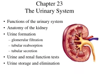
Chapter 23The Urinary System • Functions of the urinary system • Anatomy of the kidney • Urine formation • glomerular filtration • tubular reabsorption • tubular secretion • Urine and renal function tests • Urine storage and elimination
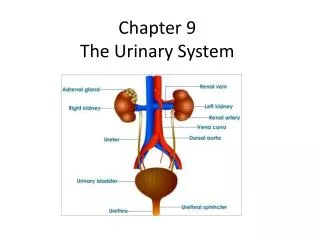
Urinary System • Two kidneys • Two ureters • Urethra
2/3s of human body is water • w/out resupply a human cannot function beyond 5/6 days • Cannot survive beyond 10 or 12 • 1 quart a day will sustain life
Why is drinking sea water lethal? • Drinking seawater is lethal within 7 – 10 days, but you’ll be conscious till the end • Blood: 1 % salt • Seawater: 3% salt • If you can make salt water less than 1% salt you can drink it. Add 1 liter of sea water to every 2 liters fresh water
Kidney Functions • Filters blood plasma, eliminates waste, returns useful substances to blood • Regulates blood volume and pressure • Regulates osmolarity of body fluids • Secretes renin (activates angiotensin II aldosterone pathway) • controls BP, electrolyte balance • Secretes erythropoietin • Regulates acid base balance • Detoxifies free radicals and drugs • Gluconeogenesis
Nitrogenous Wastes • Urea • proteinsamino acids NH2 removed forms ammonia, liver converts to urea • Uric acid • nucleic acid catabolism • Creatinine • creatine phosphate catabolism • Renal failure • azotemia: BUN, nitrogenous wastes in blood • uremia: toxic effects as wastes accumulate
Excretion • Separation of wastes from body fluids and eliminating them • respiratory system: CO2 • integumentary system: water, salts, lactic acid, urea • digestive system: water, salts, CO2, lipids, bile pigments, cholesterol • urinary system: many metabolic wastes, toxins, drugs, hormones, salts, H+ and water
Anatomy of Kidney • Position, weight and size • retroperitoneal, level of T12 to L3 • about 160 g each • about size of a bar of soap (12x6x3 cm) • Shape • lateral surface - convex; medial - concave • CT coverings • renal fascia: binds to abdominal wall • adipose capsule: cushions kidney • renal capsule: encloses kidney like cellophane wrap
Anatomy of Kidney • Renal cortex: outer 1 cm • Renal medulla: renal columns, pyramids - papilla • Lobe of kidney: pyramid and it’s overlying cortex
Kidney: Frontal Section • Minor calyx: cup over papilla collects urine
Renal Corpuscle Glomerular filtrate collects in capsular space, flows into renal tubule
Renal (Uriniferous) Tubule • Proximal convoluted tubule (PCT) • longest, most coiled, simple cuboidal with brush border • Nephron loop - U shaped; descending + ascending limbs • thick segment (simple cuboidal) initial part of descending limb and part or all of ascending limb, active transport of salts • thin segment (simple squamous) very water permeable • Distal convoluted tubule (DCT) • cuboidal, minimal microvilli
Renal (Uriniferous) Tubule 2 • Collecting duct • several DCT’s join • Flow of glomerular filtrate: • glomerular capsule PCT nephron loop DCT collecting duct papillary duct minor calyx major calyx renal pelvis ureter urinary bladder urethra
Nephrons • True proportions of nephron loops to convoluted tubules shown • Cortical nephrons (85%) • short nephron loops • efferent arterioles branch off peritubular capillaries • Juxtamedullary nephrons (15%) • very long nephron loops, maintain salt gradient, helps conserve water
Nephron Diagram • Peritubular capillaries shown only on right
Path of Blood Through Kidney • Renal artery interlobararteries(up renal columns, between lobes) arcuatearteries (over pyramids) interlobulararteries (up into cortex) afferentarterioles glomerulus (cluster of capillaries) efferentarterioles (near medulla vasa recta) peritubular capillaries interlobular veins arcuate veins interlobar veins • Renal vein
Filtration Membrane • Fenestrated endothelium • 70-90nm pores exclude blood cells • Basement membrane • proteoglycan gel, negative charge excludes molecules > 8nm • blood plasma 7% protein, glomerular filtrate 0.03% • Filtration slits • podocyte arms have pedicels with negatively charged filtration slits, allow particles < 3nm to pass
Glomerular Filtration Rate (GFR) • Filtrate formed per minute • GFR = NFP x Kf125 ml/min or 180 L/day, male • GFR = NFP x Kf105 ml/min or 150 L/day, female • filtration coefficient (Kf) depends on permeability and surface area of filtration barrier • 99% of filtrate reabsorbed, 1 to 2 L urine excreted
Effects of GFR Abnormalities • GFR, urine output rises dehydration, electrolyte depletion • GFR wastes reabsorbed (azotemia possible) • GFR controlled by adjusting glomerular blood pressure • autoregulation • sympathetic control • hormonal mechanism: renin and angiotensin
- vasomotion - monitor salinity Juxtaglomerular Apparatus
Renal Autoregulation of GFR • BP constrict afferent arteriole, dilate efferent • BP dilate afferent arteriole, constrict efferent • Stable for BP range of 80 to 170 mmHg (systolic) • Cannot compensate for extreme BP
Renal Autoregulation of GFR • Myogenic mechanism • BP stretches afferent arteriole afferent arteriole constricts restores GFR • Tubuloglomerular feedback • Macula densa on DCT monitors tubular fluid and signals juxtaglomerular cells (smooth muscle, surrounds afferent arteriole) to constrict afferent arteriole to GFR
Sympathetic Control of GFR • Strenuous exercise or acute conditions (circulatory shock) stimulate afferent arterioles to constrict • GFR and urine production, redirecting blood flow to heart, brain and skeletal muscles
-efferent arterioles Hormonal Control of GFR
Peritubular Capillaries • Blood has unusually high COP here, and BHP is only 8 mm Hg (or lower when constricted by angiotensin II); this favors reabsorption • Water absorbed by osmosis and carries other solutes with it (solvent drag)
Proximal Convoluted Tubules (PCT) • Reabsorbs 65% of GF to peritubular capillaries • Great length, prominent microvilli and abundant mitochondria for active transport • Reabsorbs greater variety of chemicals than other parts of nephron • transcellular route - through epithelial cells of PCT • paracellular route - between epithelial cells of PCT • Transport maximum: when transport proteins of cell membrane are saturated; bloodglucose > 220 mg/dL some remains in urine (glycosuria); glucose Tm = 320 mg/min
Tubular Secretion of PCT and Nephron Loop • Waste removal • urea, uric acid, bile salts, ammonia, catecholamines, many drugs • Acid-base balance • secretion of hydrogen and bicarbonate ions regulates pH of body fluids • Primary function of nephron loop • water conservation • generates salinity gradient, allows CD to conc. urine • also involved in electrolyte reabsorption
DCT and Collecting Duct • Principal cells – receptors for hormones • Intercalated cells – involved in acid/base balance • Function • BP renin release angiotensin II formation • angiotensin II stimulates adrenal cortex • adrenal cortex secretes aldosterone • promotes Na+ reabsorption promotes water reabsorption urine volumemaintainsBP
DCT and Collecting Duct 2 • Effect of ADH • dehydration stimulates hypothalamus • hypothalamus stimulates posterior pituitary • posterior pituitary releases ADH • ADH water reabsorption • urine volume
DCT and Collecting Duct 2 • Opposing effect of atrial natriuretic peptide (ANP) • BP stimulates right atrium to secrete ANP • promotes Na+ and water excretion, urine volume, blood volume • inhibits renin/angiotensin/aldosterone pathway • BP drops
DCT and Collecting Duct 2 • Effect of PTH • blood Ca2+ • calcium reabsorption • phosphate reabsorption, new bone formation • stimulates kidney production of calcitriol
Collecting Duct Concentrates Urine • Osmolarity 4x as concentrated deep in medulla • Medullary portion of CD is more permeable to water than to NaCl
Control of Water Loss • Producing hypotonic urine • NaCl reabsorbed by cortical CD • water remains in urine • Producing hypertonic urine • dehydration ADH aquaporin channels, CD’s water permeability • more water is reabsorbed • urine is more concentrated
Countercurrent Multiplier • Recaptures NaCl and returns it to renal medulla • Descending limb • reabsorbs water but not salt • concentrates tubular fluid • Ascending limb • reabsorbs Na+, K+, and Cl- • maintains high osmolarity of renal medulla • impermeable to water • tubular fluid becomes hypotonic • Recycling of urea: collecting duct-medulla • urea accounts for 40% of high osmolarity of medulla
Countercurrent Exchange System • Formed by vasa recta • provide blood supply to medulla • do not remove NaCl from medulla • Descending capillaries • water diffuses out of blood • NaCl diffuses into blood • Ascending capillaries • water diffuses into blood • NaCl diffuses out of blood
Composition and Properties of Urine • Appearance • almost colorless to deep amber; yellow color due to urochrome, from breakdown of hemoglobin (RBC’s) • Odor - as it stands bacteria degrade urea to ammonia • Specific gravity • density of urine ranges from 1.001 -1.028 • Osmolarity - (blood - 300 mOsm/L) ranges from 50 mOsm/L to 1,200 mOsm/L in dehydrated person • pH - range: 4.5 - 8.2, usually 6.0 • Chemical composition: 95% water, 5% solutes • urea, NaCl, KCl, creatinine, uric acid