Urinary System – Part I
Urinary System – Part I. Chapter 14 BIO 160 Kelly Trainor. Functions of the Urinary System. Elimination of waste products Nitrogenous wastes Toxins Drugs Regulate aspects of homeostasis Water balance Electrolytes Acid-base balance in the blood Blood pressure


Urinary System – Part I
E N D
Presentation Transcript
Urinary System – Part I Chapter 14 BIO 160 Kelly Trainor
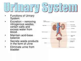
Functions of the Urinary System • Elimination of waste products • Nitrogenous wastes • Toxins • Drugs • Regulate aspects of homeostasis • Water balance • Electrolytes • Acid-base balance in the blood • Blood pressure • Red blood cell production • Activation of vitamin D
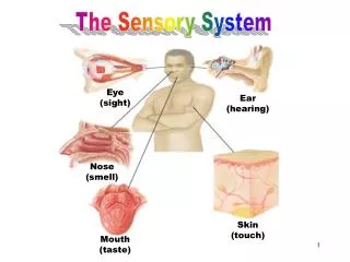
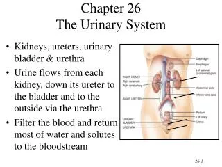
Organs of the Urinary System • Kidneys • Ureters • Urinary bladder • Urethra
Location of the Kidneys • Against the dorsal body wall • At the level of the T12 to L3 vertebrae • The right kidney is slightly lower than the left (due to position of the liver)
Kidney Features • Renal hilum • A medial indentation where several structures enter or exit the kidney (ureters, renal blood vessels, and nerves) • An adrenal gland sits atop each kidney
Coverings of the Kidneys • Fibrous capsule • Surrounds each kidney • Perirenal fat capsule • Surrounds the kidney and cushions against blows • Renal fascia • Outermost capsule that helps hold the kidney in place against the muscles of the trunk wall
Regions of the Kidney • Renal cortex—outer region • Renal medulla—inside the cortex • Renal pelvis—inner collecting tube
Kidney Structures • Renal or medullary pyramids—triangular regions of tissue in the medulla • Renal columns—extensions of cortex-like material inward that separate the pyramids • Calyces—cup-shaped structures that funnel urine towards the renal pelvis
Blood Supply • One-quarter of the total blood supply of the body passes through the kidneys each minute • Renal artery provides each kidney with arterial blood supply
Nephron Anatomy and Physiology • The structural and functional units of the kidneys • Responsible for forming urine • Main structures of the nephrons • Glomerulus • Renal tubule
Nephrons Figure 15.3a
Nephron Anatomy • Glomerulus • Knot of capillaries • Capillaries are covered with podocytes from the renal tubule • Glomerulus sits within a glomerular (Bowman’s) capsule (the first part of the renal tubule)
Glomerulus • Fed and drained by arterioles • Afferent arteriole—arises from a cortical radiate artery and feeds the glomerulus • Efferent arteriole—receives blood that has passed through the glomerulus • Specialized for filtration • High pressure forces fluid and solutes out of blood and into the glomerular capsule
Nephron Anatomy • Renal tubule extends from glomerular capsule and ends at the collecting duct • Glomerular (Bowman’s) capsule • Proximal convoluted tubule (PCT) • Loop of Henle • Distal convoluted tubule (DCT)
Nephron Anatomy Figure 15.3b
Types of Nephrons • Cortical nephrons • Located entirely in the cortex • Includes most nephrons • Juxtamedullary nephrons • Found at the boundary of the cortex and medulla
Collecting Duct • Receives urine from many nephrons • Run through the medullary pyramids • Deliver urine into the calyces and renal pelvis
Nephron Anatomy • Nephrons are associated with two capillary beds • Glomerulus • Peritubular capillary bed
Peritubular Capillary Beds • Arise from efferent arteriole of the glomerulus • Normal, low pressure capillaries • Adapted for absorption instead of filtration • Cling close to the renal tubule to reabsorb (reclaim) some substances from collecting tubes
Urine Formation • Glomerular filtration • Tubular reabsorption • Tubular secretion
Glomerular Filtration • Nonselective passive process • Water and solutes smaller than proteins are forced through capillary walls • Proteins and blood cells are normally too large to pass through the filtration membrane • Filtrate is collected in the glomerular capsule and leaves via the renal tubule
Tubular Reabsorption • The peritubular capillaries reabsorb useful substances • Water • Glucose • Amino acids • Ions • Some reabsorption is passive, most is active • Most reabsorption occurs in the proximal convoluted tubule • Materials not reabsorbed • Nitrogenous waste products • Urea—protein breakdown • Uric acid—nucleic acid breakdown • Creatinine—associated with creatine metabolism in muscles
Tubular Secretion • Some materials move from the peritubular capillaries into the renal tubules • Hydrogen and potassium ions • Creatinine • Process is important for getting rid of substances not already in the filtrate • Materials left in the renal tubule move toward the ureter
Characteristics of Urine • In 24 hours, about 1.0 to 1.8 liters of urine are produced • Urine and filtrate are different • Filtrate contains everything that blood plasma does (except proteins) • Urine is what remains after the filtrate has lost most of its water, nutrients, and necessary ions • Urine contains nitrogenous wastes and substances that are not needed
Characteristics of Urine • Yellow color due to the pigment urochrome (from the destruction of hemoglobin) and solutes • Sterile • Slightly aromatic • Normal pH of around 6 • Specific gravity of 1.001 to 1.035
Characteristics of Urine • Solutes normally found in urine • Sodium and potassium ions • Urea, uric acid, creatinine • Ammonia • Bicarbonate ions • Solutes NOT normally found in urine • Glucose • Blood proteins • Red blood cells • Hemoglobin • White blood cells (pus) • Bile
Urinary System – Part II Chapter 14 BIO 160 Kelly Trainor
Ureters • Slender tubes attaching the kidney to the bladder • Continuous with the renal pelvis • Enter the posterior aspect of the bladder • Runs behind the peritoneum • Peristalsis aids gravity in urine transport
Organs of the Urinary System Figure 15.1a
Urinary Bladder • A moderately full bladder is about 5 inches long and holds about 500 mL of urine • Capable of holding twice that amount of urine • Smooth, collapsible, muscular sac • Temporarily stores urine • Trigone—triangular region of the bladder base • Three openings • Two from the ureters • One to the urethra • In males, the prostate gland surrounds the neck of the bladder
Female Urinary Bladder and Urethra Figure 15.6
Urinary Bladder Wall • Three layers of smooth muscle collectively called the detrusor muscle • Mucosa made of transitional epithelium • Walls are thick and folded in an empty bladder • Bladder can expand significantly without increasing internal pressure
Urethra • Thin-walled tube that carries urine from the bladder to the outside of the body by peristalsis • Release of urine is controlled by two sphincters • Internal urethral sphincter • Involuntary and made of smooth muscle • External urethral sphincter • Voluntary and made of skeletal muscle
Urethra Gender Differences • Length • Females is 3–4 cm (1 inch) • Males is 20 cm (8 inches) • Location • Females—along wall of the vagina • Males—through the prostate and penis • Function • Females—only carries urine • Males—carries urine and is a passageway for sperm cells
Micturition (Voiding) • Both sphincter muscles must open to allow voiding • The internal urethral sphincter is relaxed after stretching of the bladder • Pelvic splanchnic nerves initiate bladder to go into reflex contractions • Urine is forced past the internal urethra sphincter and the person feels the urge to void • The external urethral sphincter must be voluntarily relaxed to void
Urinary System – Part III Chapter 14 BIO 160 Kelly Trainor
Fluid, Electrolyte, and Acid-Base Balance • Blood composition depends on three factors • Diet • Cellular metabolism • Urine output • Kidneys have four roles in maintaining blood composition • Excretion of nitrogen-containing wastes (previously discussed) • Maintaining water balance of the blood • Maintaining electrolyte balance of the blood • Ensuring proper blood pH
Distribution of Body Fluid • Intracellular fluid (ICF) • Fluid inside cells • About two-thirds of body fluid • Extracellular fluid (ECF) • Fluids outside cells that includes • Interstitial fluid • Blood plasma
The Link Between Water and Salt • Solutes in the body include electrolytes like sodium, potassium, and calcium ions • Changes in electrolyte balance causes water to move from one compartment to another • Alters blood volume and blood pressure • Can impair the activity of cells
Maintaining Water Balance • Water intake must equal water output • Sources for water intake • Ingested foods and fluids • Water produced from metabolic processes • Thirst mechanism is the driving force for water intake • Sources for water output • Vaporization out of the lungs • Lost in perspiration • Leaves the body in the feces • Urine production
Maintaining Water Balance • Dilute urine is produced if water intake is excessive • Less urine (concentrated) is produced if large amounts of water are lost • Proper concentrations of various electrolytes must be present
Maintaining Acid-Base Balance in Blood • Blood pH must remain between 7.35 and 7.45 to maintain homeostasis • Alkalosis—pH above 7.45 • Acidosis—pH below 7.35 • Physiological acidosis—pH between 7.35 and 7.0 • Most ions originate as by-products of cellular metabolism
Blood Buffers • Molecules react to prevent dramatic changes in hydrogen ion (H+) concentrations • Bind to H+ when pH drops • Release H+ when pH rises • Three major chemical buffer systems • Bicarbonate buffer system • Phosphate buffer system • Protein buffer system
Renal Mechanisms of Acid-Base Balance • When blood pH rises • Bicarbonate ions are excreted • Hydrogen ions are retained by kidney tubules • When blood pH falls • Bicarbonate ions are reabsorbed • Hydrogen ions are secreted • Urine pH varies from 4.5 to 8.0