
Exploring Medical Imaging Detectors: Commonalities with CERN Technologies
E N D
Presentation Transcript
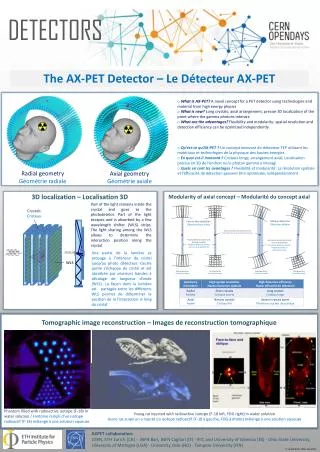
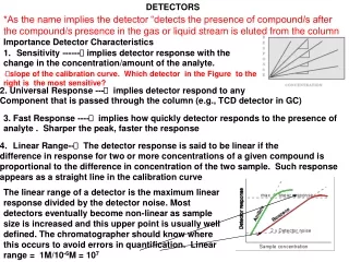
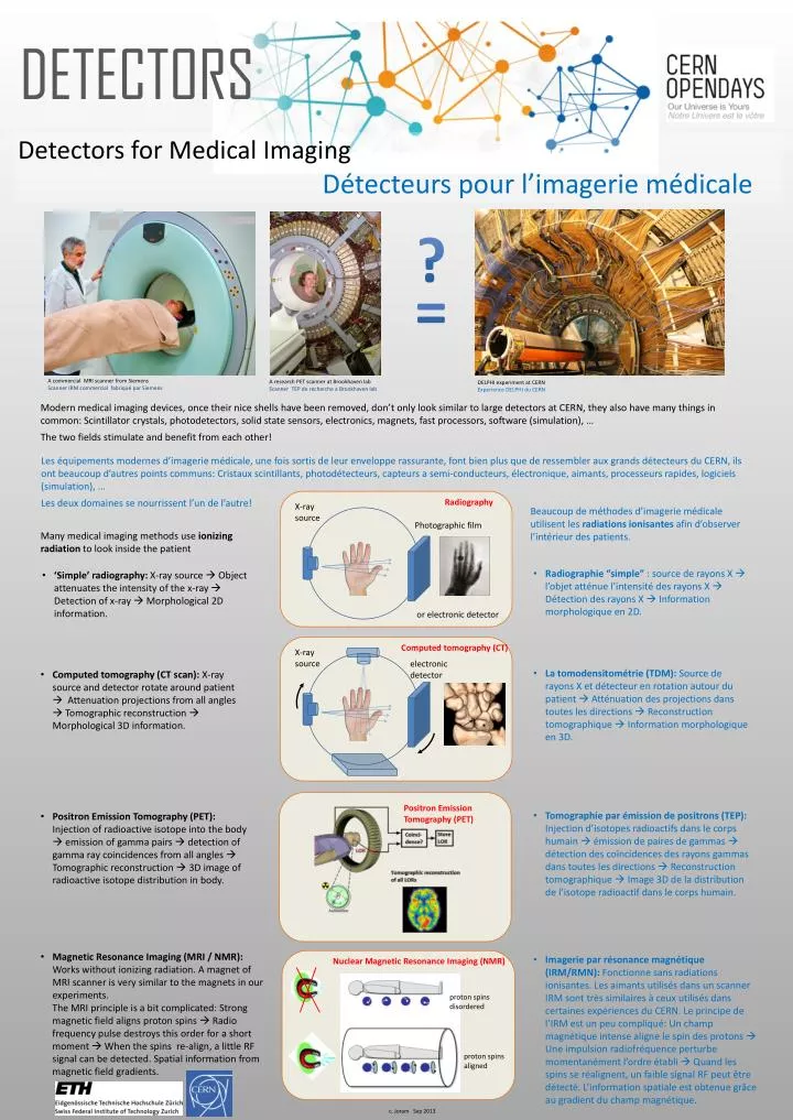
DETECTORS Detectors for Medical Imaging Détecteurspour l’imageriemédicale ? = A commercial MRI scanner from Siemens Scanner IRM commercial fabriquépar Siemens A research PET scanner at Brookhaven lab Scanner TEP de recherche a Brookhaven lab • DELPHI experiment at CERN • Experience DELPHI du CERN • Modern medical imaging devices, once their nice shells have been removed, don’t only look similar to large detectors at CERN, they also have many things in common: Scintillator crystals, photodetectors, solid state sensors, electronics, magnets, fast processors, software (simulation), … • The two fields stimulate and benefit from each other! Les équipementsmodernesd’imageriemédicale, unefoissortis de leurenvelopperassurante, font bien plus que de ressembler aux grandsdétecteursdu CERN, ilsont beaucoup d’autres points communs: Cristauxscintillants, photodétecteurs, capteurs a semi-conducteurs, électronique, aimants, processeursrapides, logiciels(simulation), … Les deuxdomaines se nourrissentl’un de l’autre! Radiography X-ray source Photographic film or electronic detector • Beaucoup de méthodesd’imageriemédicaleutilisent les radiations ionisantesafind’observerl’intérieur des patients. • Many medical imaging methods use ionizing radiation to look inside the patient • Radiographie “simple” : source de rayons X l’objetatténuel’intensité des rayons X Détection des rayons X Information morphologique en 2D. • ‘Simple’ radiography:X-ray source Object attenuates the intensity of the x-ray Detection of x-ray Morphological 2D information. Computed tomography (CT) X-ray source electronic detector • La tomodensitométrie (TDM): Source de rayons X et détecteur en rotation autour du patient Atténuation des projections danstoutes les directions Reconstruction tomographique Information morphologique en 3D. • Computed tomography (CT scan):X-ray source and detector rotate around patient Attenuation projections from all angles Tomographic reconstruction Morphological 3D information. Positron Emission Tomography (PET) • Tomographie par émission de positrons (TEP): Injection d’isotopesradioactifsdans le corps humainémission de paires de gammas détection des coïncidences des rayons gammas danstoutes les directions Reconstruction tomographique Image 3D de la distribution de l’isotoperadioactifdans le corps humain. • Positron Emission Tomography (PET): Injection of radioactive isotope into the body emission of gamma pairs detection of gamma ray coincidences from all angles Tomographic reconstruction 3D image of radioactive isotope distribution in body. • Magnetic Resonance Imaging (MRI / NMR): Works without ionizing radiation. A magnet of MRI scanner is very similar to the magnets in our experiments. • The MRI principle is a bit complicated: Strong magnetic field aligns proton spins Radio frequency pulse destroys this order for a short moment When the spins re-align, a little RF signal can be detected. Spatial information from magnetic field gradients. • Imagerie par résonancemagnétique (IRM/RMN): Fonctionne sans radiations ionisantes. Les aimantsutilisésdans un scanner IRM sonttrèssimilaires à ceuxutilisésdanscertainesexpériences du CERN. Le principe de l’IRMest un peucompliqué: Un champ magnétique intense aligne le spin des protons Une impulsion radiofréquenceperturbemomentanémentl’ordreétabliQuand les spins se réalignent, un faible signal RF peutêtredétecté. L’informationspatialeestobtenue grâce au gradient du champ magnétique. Nuclear Magnetic Resonance Imaging (NMR) proton spins disordered proton spins aligned c. Joram Sep 2013