Download
1 / 87
931 likes | 1.69k Views
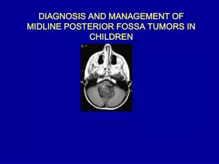
DIAGNOSIS AND MANAGEMENT OF MIDLINE POSTERIOR FOSSA TUMORS IN CHILDREN. INTRODUCTION. Medulloblastoma Ependymoma Astrocytoma Brainstem glioma Choroid plexus papilloma Dermoid. Me dulloblastoma. Bailey and Cushing in 1925 first used the term medulloblastoma.

E N D
DIAGNOSIS AND MANAGEMENT OF MIDLINE POSTERIOR FOSSA TUMORS IN CHILDREN
INTRODUCTION Medulloblastoma Ependymoma Astrocytoma Brainstem glioma Choroid plexus papilloma Dermoid
Medulloblastoma Bailey and Cushing in 1925 first used the term medulloblastoma. One of the most common tumors of posterior fossa (20 – 25 % all pediatric brain tumors) 5 –7 yrs – median age of diagnosis. 2 – 4 and 6 –8 yrs : two peaks in children
Medulloblastoma Histologic subtypes: Classical medulloblatoma Desmoplastic medulloblastoma Medullomyoblastoma Melanotic medulloblastoma Large-cell medulloblastoma: Very poor outcome
Medulloblastoma….origin Debatable: Origin from remnant of cells of the external granular layer of the cerebellum. Transformation of normal undifferentiated progenitor cells of superior medullary velum which migrate to the external granular layer.
Medulloblastoma….Clinical Hydrocephalus : Raised ICP Behavioral change, listlessness, irritability, vomiting, and decreased social interactions. Headache, especially in the morning. Double vision. Head tilt : tonsillar herniation below the foramen magnum. (Can result from trochlear nerve palsy caused by direct tumor compression )
Medulloblastoma….Clinical Cerebellar symptoms Brain stem involvement Leptomeningeal dissemination
Medulloblastoma….Clinical Physical: Increasing head circumference , full anterior fontanalles with widely split cranial sutures. Fundus examination Papilledema can be present in as many as 90% of patients.
Medulloblastoma….Clinical Extraocular examination Diplopia and lateral gaze paresis Fourth cranial nerve palsy ( should be considered in any patient with a head tilt ) Nystagmus Cerebellar signs ( ataxia > unilateral dysmetria )
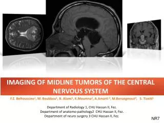
Radiology…….CT CECT NCCT
Radiology…….MRI Homogeneous enhancement ( may be absent in about 15 – 20 % ) DWI shows restricted diffusion with increased ADC. MRI spine : Should be done at time of diagnosis. BEST : prior to surgery. If not possible Should be delayed for at least 2 weeks after surgery.
Radiology……. Skeletal imaging Metastasis to the bone must be considered in any child with medulloblastoma and bone pain. A skeletal survey helps elucidate lytic or sclerotic lesions.
Diagnosis …..CSF cytology No standardized method: HOW and WHEN ?? Lumbar puncture Ventricular drain Cisterna magna at the time of surgery from the for cytologic analysis.
Staging…….. Within 48 hours of surgery, a Gd MRI. Staging. Assess residual tumor size prior to the onset of enhancing reactive gliosis. Staging is dependent upon : extent of resection, radiographic evidence of tumor spread, and CSF cytology.
Current staging of medulloblastoma Standard Risk Posterior fossa No metastasis < 1.5 cm2 residual Undifferentiated High Risk Posterior fossa with intracranial or spinal dissemination. Extra neural metastasis > 1.5 cm2 residual Differentiated
Diagnosis…..genetics Routine use : Controversial. Correlation between aneuploid DNA content and a better prognosis. 17qi an isochromosome : Most common C-ERB2 –poor outcome Neurotropin growth factor receptor (TrkC) expression: associated with better outcome.
Risk factors associated with outcome for medulloblastoma Good Prognosis Females Sex Gross total resection No metastasis Desmoplastic histology Increased apoptosis index Hyperdiploidy High TRKC expression Poor Prognosis Younger age Subtotal resection Metastasis Large-cell anaplastic histology Elevated Ki-67/MIB index Aneuploidy Elevated ERB2 expression Isolated 17p LOH Elevated expression and amplification of MYCC Up regulation of PDGFR Over expression of calbindin- D28k Fisher PG etal. Biologic Risk Stratification of Medulloblastoma: The Real Time Is Now. J Clin Oncol 2004;22; 971-74
Presenation : MRI Brain and spine Surgical resection Management of hydrocephalus < 3 years > 3 years Standard risk Poor risk Chemotherapy (No standard regimen) Craniospinal radiation OR Reduced dose radiation with CT on reasarch protocol Craniospinal radiation + adjunct CT ( CCNU, cisplatin vincristine or CT on research protocol Follow OR Delayed RT till 3 years old Management algorithm for medulloblastoma
Hydrocephalus The majority of children with posterior fossa tumors have hydrocephalus at the time of presentation. There is no consensus regarding the management of HC in these children
Hydrocephalus Treatment options: Ventriculoperitoneal shunt Perioperative EVD Endoscopic third ventriculostomy Direct surgical resection
Hydrocephalus………. Recent studies have shown that ultimately 17 to 40% of children have uncontrolled hydrocephalus and require shunt placement during the postoperative period; and that this predominantly occurred within the 1st postoperative month. An expectant policy in these subgroup who ultimately require a shunt place them at risk of developing intracranial hypertension ,an increased rate of CSF leakage, and pseudomeningocele formation, prolonged hospitalization.
Hydrocephalus …….…Factors predicting patients at risk of requiring placement of a shunt postoperatively Younger age at diagnosis The severity of hydrocephalus prior to resection of the tumor Midline localization Incomplete tumor removal Use of substitute dural grafts during closure CSF infection Persistent pseudomeningocele An analysis of factors determining the need for ventriculoperitoneal shunts after posterior fossa tumor surgery in children. Neurosurgery 34:402-408, 1994 Pediatr Neurosurg 20:240-247, 1994
Management…….. Surgery Gross Total Resection, if possible. Brainstem damage should be avoided. Resolution of natural CSF pathways. Tumor adheres to the floor of the fourth ventricle, precluding gross total resection.( 1/3 rd of cases ) Sugar coating – subarachnoid spread.
Management…….. Radiotherapy SURGERY alone : NOT CURATIVE RADIOTHERAPY : cornerstone of adjuvant therapy. 54 to 58 Gy to the primary site with 35Gy to the entire craniospinal axis Institution of presymptomatic craniospinal radiation therapy is probably the single most important factor responsible for the improved survival rates
Management…….. Radiotherapy Complications of radiotherapy : lowered intelligence quotient (IQ), small stature, endocrine dysfunction, behavioral abnormalities, secondary neoplasms white matter necrosis. Reduction in IQ and neurobehavioral function.
Management….. Hyperfractionated radiotherapy Delivery of higher doses of radiation without increased toxicity. The typical hyperfractionated radiotherapy schedule consists of twice-daily fraction sizes of 100 to 120 cGy to a total dose of 7200 to 7800 cGy. In practice hyperfractionated therapy has shown no advantage over the standard RT.
Management……. Chemotherapy EXACT BENEFITS : UNCLEAR Delay the onset of radiation therapy in young children ( < 3 years ) Increase in survival rates in high-risk children with medulloblastoma Patients with recurrent or advanced disease Reduction in the RT dose to the neuraxis in patients with nondisseminated disease
Management…….. New studies Sensitizing the tumor to irradiation with the concomitant use of chemotherapy. Presurgical chemotherapy to treat patients prior to surgery. Intraventricular administration of cytotoxic agents, Newer drug combinations, and Immunotherapy based on genetics analysis
Management…….. Recurrent Medulloblastoma Recurrences : 30 to 40% of patients Chemotherapy : limited due to chemo resistance in those patients who have previously undergone CT Redosing with RT avoided due to radiation necrosis. ( Local RT using stereotactic techniques can be used can palliative )
Management…….. Recurrent Medulloblastoma High-dose chemotherapy with autologous SCR or autologous BMR : subject of intense investigation. Stem cell rescue involves harvesting autologous bone marrow or preferably, peripheral stem cells by using pheresis techniques and subsequently reinfusing them after provision of high-dose myeloablative chemotherapy. Int J Legal Med. 2001;114(6):331-7 • Substantial toxicity : • Death, serious infection, and venoocclusive disease.
Management…….. Recurrent Medulloblastoma Though data suggests longer EFS. ( In the absence of RCT, the interpretation of the results remains limited ) Benefits seen in a subset of patients, with locally recurrent disease (not involving the brainstem) and without evidence of dissemination.
Management…….. Prognosis 5 - year recurrence-free survival rates : 55% - 67%. Even after a good response to surgery and radiation, recurrence is common. Most common site : PRIMARY TUMOR SITE Bone : most common site of systemic metastasis; followed by regional lymph node.
AIIMS Protocol Surgical resection Management of hydrocephalus CSF +VE CSF - VE Cranial RT – 56Gy / 30# / 6 wks. ( 36 Gy/20# followed by a boost of 20Gy /10 # ) Spinal RT – 30 Gy / 20# / 4 wks. Concurrently with cranial RT) Dose of spinal RT 36Gy/30#/6 weeks
Cerebellar Mutism Cerebellar mutism was first reported in 1979 by Hirsh after a posterior fossa tumor resection. Also known as posterior fossa syndrome Approximately 10 -1 5 % of children undergoing posterior fossa surgery for tumor.
Cerebellar Mutism Decreased or absent speech, irritability, hypotonia, ataxia. Onset : Immediate or delayed. Virtually all cases of mutism will occur within the first week of surgery ( 50% within the first two days ) Most cases resolves in a week or two.( longest 52 months) with return of functional speech.
Factors associated with the development of mutism Posterior fossa surgery for tumor. Children Midline tumor location Cerebellar vermal incision Large tumor size ( > 5cm ) Medulloblastoma
Cerebellar Mutism…. Pathophysiology. UNKNOWN. However not emotional. Focal decreased cerebral and cerebellar blood flow leading to decreased cell functioning in particular areas, dentate-thalami-cortical pathway causing dysfunction. SPECT studies have lead support to this theory
Cerebellar Mutism…. Outcome Speech almost always returns. The speech is virtually always becomes functional for communication, however it may not be the same as before surgery.
Cerebellar Mutism…. intervention Speech therapy Assisting in some form of nonverbal communication Reassurance : usual course of cerebellar mutism and what to expect in the recovery. Practicing tongue and lip movements before speech returns
Brain Stem Gliomas Brainstem tumors comprise 10–20% of all pediatric central nervous system tumors. Once considered uniformly fatal ; the perspective has changed now.
Clinical hallmark Bilateral long tract signs Bilateral multiple contiguous cranial nerve palsies. Horner’s syndrome Inter Nuclear Ophthalmoplegia
BSG……Classification The most recent classification system by Choux et al based on both CT and MRI imaging Type I – Diffuse Type II – Intrinsic, focal Type III – Exophytic, focal Type IV – Cervicomedullary Pediatric Neurosurgery. New York, Churchill Livingstone, 2000, pp 471–491.
BSG…… Type I : Diffuse brainstem gliomas Appro. 75% of all tumors Hypointense on CT No significant enhancement on MRI. Characterized by diffuse infiltration and swelling of the brainstem. Typically, are malignant fibrillary astrocytomas (WHO grade III or IV).
BSG…… Type II : Focalintrinsic tumors ( cystic/solid ) Sharply demarcated from surrounding tissue on MRI and are associated with less brainstem edema. Majority of these lesions are low grade gliomas (WHO I or II). Contrast enhancement : variable
BSG…… Type III : Exophytic tumors that arise from the subependymal glial tissue of the fourth ventricle and mostly grow dorsally or laterally. MRI characteristics similar to type II lesions, and histologically, these lesions are usually low-grade lesions (WHO I or II) like type II lesions.