Download

1 / 61
670 likes | 1.17k Views
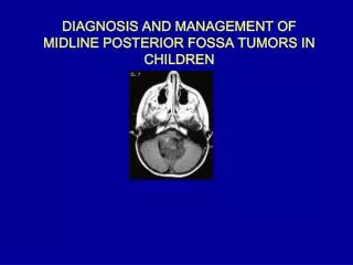
POST FOSSA TUMORS DIAGNOSIS AND TREATMENT. Introduction. Primary brain tumor – 6 persons/100000/year Metastatic brain tumor – 6 persons/100000/year 1 in 15 primary brain tumors occur in children under 15 years.

E N D
Introduction Primary brain tumor – 6 persons/100000/year Metastatic brain tumor – 6 persons/100000/year 1 in 15 primary brain tumors occur in children under 15 years In adults, the commonest tumors are gliomas, metastases and meniongiomas; most lie in the supratentorial compartment
Intra-axial Post Fossa Tumors Paediatric • PNET (including medulloblastoma) 27% • Cerebellar (Pilocyticastrocytoma) 27% • Brain stem glioma 27% • Ependymoma 15% • Choroid plexus papilloma (<1% of primary brain tumor) • Dermoid cyst (<0.5% of primary intraaxial tumor) • Atypical teratoid/ rhabdoid tumor Adult • Metastasis 16% • Hemangioblastoma 7-12% • Pilocytic astrocytoma (2nd decade) • Brain stem glioma (1% of adult tumor) • Choroid plexus tumor (<1% of primary brain tumor) • Cerebellar liponeurocytoma
Extra-axial Lesion • Vestibular schwannoma • Meningioma • Epidermoid • Metastases • Trigeminal neuroma • Facial nerve neuroma • Arachnoid cyst
Metastases • Cerebellum is a common site • 16% of cases of solitary brain mets • MC post fossa tumor in adults Primary • Lung – 44% • Breast – 10% • Kidney (renal cell) – 7% • GI – 6% • Melanoma – 3% • Undetermined – 10%
Pathology • Rounded solid partially cystic mass ± edema Age • Rare in children, most common in older adults (> 40 years) Location • Anywhere: grey white junction most common site Imaging • NECT: Iso / hyperdense; Ca++ rare in untreated metastases • CECT: Strong solid/ring enhancement • MR: Most hypointense on T1, hyperintense on T2W1, most enhance moderately intensely following contrast administration
Management • Mostly palliative • Median survival of patient 26-32 weeks Medical • Corticosteroids • Anticonvulsants.
Surgical Management Solitary lesion Surgical excision of solitary lesion: • Primary disease quiescent or radioresistant • Lesion accessible, symptomatic or life threatening • For recurrent small cell lung carcinoma following XRT • Diagnosis unknown
Multiple Lesions • Worse prognosis than solitary lesion • Usually treated with XRT without surgery Situations where surgery is done: • One particular and accessible lesion symptomatic and/or life threatening • Multiple lesions that can all be completely removed Stereotactic Biopsy • Lesions not appropriate for surgery • Not candidates for surgical resection • To ascertain a diagnosis
Stereotactic Radiosurgery • No mass effect, no hydrocephalus • Advantage: No risk of hemorrhage, infection or mechanical spread of tumor cells, Can be used for 3 or fewer mets • Disadvantage: Histological proof not obtained, Cannot be used for lesion > 3 cm
Median survival following craniotomy Median survival even with best treatment is only – 8 months
Hemangioblastoma (HGB) • Most common primary intra-axial posterior fossa tumor in adults (7-12% of post fossa tumors) • Highly vascular well circumscribed solid or cystic neoplasm of CNS or retina • May occur sporadically (4th Decade) or as part of Von Hippel Lindau disease (3rd decade) • 30% of patients with cerebellar HGB have VHL
Pathology Location • 60% cystic with nodule – 40% solid • Gross hemorrhage, calcification necrosis rare • 80% to 85% cerebellum • 3% to 13% spinal cord • 2% to 3% Medulla Supratentorial lesions occur but are uncommon • 60% of patients with VHL have retinal lesions Age • Adults with peak during 40 to 60 years, rare in children
Imaging • Vertebral Angiography: Vascular nodule with intense, prolonged stain ± avascular cyst • CT: Low density cyst with strongly enhancing mural nodule that abuts a pial surface • MR: Cyst slightly hyperintense to CSF on T1W1; hyperintense to brain on T2W1; mural nodule variable but enhances strongly Labs • Polycythemia • Catecholamine production from pheochromocytoma
Treatment • May be curative in cases of HGB, not in VHL • Preop embolisation reduces the vascularity • Cystic hemagloblastoma require removal of mural nodule. Stereotactic Radiosurgery • For asymptomatic HGB > 5 mm diameter if they are cystic or progressing in size during surveillance
Radiation Treatment • Effectiveness dubious • May be useful to reduce tumor size or to retard growth in patients who are not surgical candidates for multiple brainstem HGB Chemotherapy • Ongoing phase II trial with Sunitnib, an inhibitor of vascular endothelial growth factor and platelet derived growth factor
Medulloblastoma Origin of cells (WHO- PNET) Static- external granular layer Origin from remnant of cells of the external granular layer of the cerebellum. Dynamic – neural progenitor cells Transformation of normal undifferentiated progenitor cells of superior medullary velum which migrate to the fourth ventricle
Medulloblastoma Histology • Medulloblastoma (Grade 4) • Desmoplastic/nodular medulloblastoma • Medulloblastoma with extensive nodularity • Anaplastic medulloblastoma • Large cell medulloblastoma
Medulloblastoma • Histology Cellular, small cells, scant cytoplasm, Homer-Wright rosettes Immuno histochemistry GFAP + EMA –
Medulloblastoma Clinical features Hydrocephalus : Raised ICP • Behavioral change, listlessness, irritability, vomiting, and decreased social interactions. • Headache • Double vision. • Head tilt : tonsillarherniation below the foramen magnum • Cerebellar symptoms • Brain stem involvement • Leptomeningeal dissemination
Medulloblastoma Examination • Increasing head circumference , full anterior fontanelle with widely split cranial sutures. • Papilledema 90% of patients • Diplopia and lateral gaze paresis • Fourth cranial nerve palsy ( should be considered in any patient with a head tilt ) • Nystagmus • Cerebellar signs ( ataxia > unilateral dysmetria )
Medulloblastoma • MRI- T1- low to isointense T2- hyperintense • Homogenous contrast enhancement (may be absent in about 15 –20 % ) • DWI shows restricted diffusion with increased ADC. Spinal imaging – • At diagnosis (11-71% show dissemination) • Within 24 hrs after surgery or 2 weeks post surgery • Surveillance imaging at 3-6 months
Medulloblastoma • Management Steroids CSF cytology- LP, EVD, Cisternamagna CSF diversion Definitive surgery Adjuvant therapy
CHANG CLASSIFICATION Medulloblastoma Stage Feature Tumor stage T1 Less than 3 cm diameter, limited to vermis, roof of fourth ventricle, or hemisphere T2 More than 3 cm diameter, invades one adjacent structure or partially fills fourth ventricle. T3a Invades two adjacent structure or completely fills fourth ventricle with extension into cerebral aqueduct, foramen of Luschka, or formen of Magndie. T3b Arises from floor of fourth ventricle or brain stem; fourth ventricle completely filledT4 Spreads to involve cerebral aqueduct, third ventrical, midbrain, or upper cervical spinal cord Metastasis stageM0 No evidence of metastasis M1 Tumor cells in CSF M2 Gross nodular seeding of brain CSF spaces M3 Gross nodular seeding of spinal CSF spaces M4 Extraneural spread
Current staging of medulloblastoma • HIGH RISK • Bulky residual tumor > 1.5 cm2 postop • Dissemination in the brain, spine or CSF • Worse prognosis • 5 year disease free survival is 35-50% STANDARD RISK • No residual tumor on postop MRI and negative CSF result • 5 years survival is >5% and progression free survival = 50%
Presenation : MRI Brain and spine Surgical resection Management of hydrocephalus < 3 years > 3 years Chemotherapy (No standard regimen) Standard risk Poor risk Craniospinal radiation OR Reduced dose radiation with CT on reasarch protocol Cranispinal radiation + adjunct CT ( CCNU, cisplatin vincristine or CT on research protocol Follow OR Delayed RT till 3 years old Management algorithm for medulloblastoma
Management…….. Surgery • Gross Total Resection, if possible (arises from roof of fourth ventricle- soft reddish vascular with some times sugar coating). • Brainstem damage should be avoided. • Resolution of natural CSF pathways. • SURGERY alone : NOT CURATIVE • RADIOTHERAPY : Cornerstone of adjuvant therapy. • 54 to 58 Gy - primary site • 35Gy - craniospinal axis
Management…….. Recurrent Medulloblastoma • Chemotherapy : limited due to chemo resistance in those patients who have previously undergone CT • Redosing with RT avoided due to radiation necrosis • High-dose chemotherapy with autologous SCR or autologous BMR: subject of intense investigation Prognosis • 5 - year recurrence-free survival rates : 55% - 67%. • Most common site : PRIMARY TUMOR SITE
Ependymoma • 10% of brain tumors in children • Peak age - 0-4yrs • Male preponderance • Children 90% in cranium • Adults in spinal
EPENDYMOMA • MYXOPAPILLARY (WHO Grade 1) • SUBEPENDYMOMA (WHO Grade 1) • Ependymoma (WHO Grade 2) • Cellular • Papillary • Clear cell • Tanycytic • Anaplastic ependymoma (WHO Grade 3)
Ependymoma …….. Imaging CT : Typically isodense with heterogenous enhancement Calcification : common ( can be seen in one half of cases)
Ependymoma…..MRI • On MRI, heterogeneous secondary to necrosis, hemorrhage and calcification. • Heterogenous contrast enhancement • Plasticity • Extension to the cerebellopontine angle is characteristic of ependymomas
Ependymoma….. • INTRA OP- Tumor arises from the floor and is greyishlobulated gritty and firm • Staging: No conventional staging criteria. • Postoperative MRI is recommended within 48 hours
Ependymoma…Role of Radiotherapy • Post-operative radiation recommended for patients older than 3 years. • Stereotactic radiosurgery : Therapeutic option in patients with residual, unresectable or recurrent tumor Role of Chemotherapy • May be useful < 3 years : Delay cranial radiation • Childhood intracranial ependymomas : in general chemo-resistant
AIIMS Protocol Low Grade CSF -VE High grade CSF + VE Surgery Surgery Radiotherapy 56Gy / 28# / 5.5 wks (50 Gy followed by a boost of 6 Gy) Surgery followed by CSI and 6 cycles chemotherapy.
Cerebellar (Pilocytic astrocytoma) • 10-20% of pediatric brain tumour • Pilocyticastrocytoma is the most common pediatric central nervous system glial neoplasm • Benign : extremely high survival rate 94% at 10 years • Most patients present in the first 2 decades
Pilocytic astrocytoma….MRI Four predominant imaging patterns : Mass with a nonenhancing cyst and an intensely enhancing mural nodule (21%) Mass with an enhancing cyst wall and an intensely enhancing mural nodule (46%) Necrotic mass with a central nonenhancing zone (16%), and Predominantly solid mass with minimal to no cyst like component (17%)
Pilocytic astrocytoma…. • Surgical resection of cerebellar pilocytic astrocytomas is considered the treatment of choice • Resection of mural nodule – key surgical objective • Resection of cyst wall – controversial ?? • Radiation therapy is strictly avoided, given its risk of causing significant morbidity in children younger than 5 years of age
Brainstem gliomas (BSG) • 75% in children, 25 % in adults • Median-6.5years • 1 % of pediatric brain tumors and 25 % of pediatric post fossa tumors • 75% diffuse variety • Either very benign or malignant
HALLMARKS OF BSG • Bilateral long tract signs • Bilateral multiple contiguous cranial nerve palsies • Horner’s syndrome • Inter Nuclear Ophthalmoplegia
BSG……Classification • The most recent classification system by Choux et al based on both CT and MRI imaging • Type I – Diffuse • Type II – Intrinsic, focal • Type III – Exophytic, focal • Type IV – Cervicomedullary • Pediatric Neurosurgery. New York, Churchill Livingstone, 2000, pp 471–491.
BSG…… • Type I : Diffuse brainstem gliomas • 75% of all BSG • Hypointense on CT • No significant enhancement on MRI. • Characterized by diffuse infiltration and • swelling of the brainstem. • Typically, are malignant fibrillary • astrocytomas (WHO grade III or IV).
BSG…… • Type II : Focalintrinsic tumors ( cystic/solid ) • Sharply demarcated from surrounding tissue on MRI and are associated with less brainstem edema. • Majority of these lesions are low grade gliomas (WHO I or II). • Contrast enhancement : variable
BSG…… • Type III : Exophytic tumors that arise from the subependymal glial tissue of the fourth ventricle and mostly grow dorsally or laterally. • MRI characteristics similar to type II lesions, and histologically, these lesions are usually low-grade lesions (WHO I or II) like type II lesions.
BSG…… • Type IV lesions are cervicomedullary brainstem gliomas. • Imaging, histology and behavior : similar to intramedullary spinal cord gliomas. • Majority are low-grade, non-infiltrative tumors.
BSG…..Management • Biopsy : only for indeterminate lesions • Stereotactic biopsy: can provide diagnostic tissue. • Stereotactic radiosurgery • Not without risk: Damage to the cranial nerves and long tracts Tissue heterogeneity
Management • Focal cystic tumors- SX+RT • Focal solid tumors- SX • Dorsal Exophytic tumors- SX + Focal RT • Dorsal Exophytic malignant tumor- RT+CT • Diffuse infiltrating – RT + steroids
Choroid Plexus Tumors • Neoplasms of the choroid plexus. • Lateral ventricles : most common location in children. • 4th ventricle : most common location in adults. • 4-6% of the intracranial neoplasms in children younger than 2 years. • Choroid plexus tumors • Choroid plexus papilloma (WHO Grade 1) • Atypical choroid plexus papilloma (WHO Grade 2) • Choroid plexus carcinoma (WHO Grade 3)
Choroid Plexus TUMORS…..Clinical • Hydrocephalus and raised ICT • The tumor itself can cause mass effect. • Surgery may not resolve HCP (derangement of reabsorption mechanisms or blockage at other sites in the ventricular system)