Download
1 / 14
190 likes | 649 Views
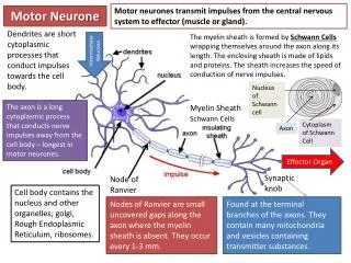
Motor neurones transmit impulses from the central nervous system to effector (muscle or gland). . Motor Neurone. Dendrites are short cytoplasmic processes that conduct impulses towards the cell body.

E N D
Motor neurones transmit impulses from the central nervous system to effector (muscle or gland). Motor Neurone Dendrites are short cytoplasmic processes that conduct impulses towards the cell body. The myelin sheath is formed by Schwann Cells wrapping themselves around the axon along its length. The enclosing sheath is made of lipids and proteins. The sheath increases the speed of conduction of nerve impulses. Intermediate Neurone Nucleus of Schwann cell The axon is a long cytoplasmic process that conducts nerve impulses away from the cell body – longest in motor neurones. Myelin Sheath Schwann Cells Cytoplasm of Schwann Cell Axon Effector Organ Synaptic knob Node of Ranvier Cell body contains the nucleus and other organelles; golgi, Rough Endoplasmic Reticulum, ribosomes. Nodes of Ranvier are small uncovered gaps along the axon where the myelin sheath is absent. They occur every 1-3 mm. Found at the terminal branches of the axons. They contain many mitochondria and vesicles containing transmitter substances.
Sensory Neurone Transmits impulses from a receptor to the CNS. Dendrons are long cytoplasmic processes that conducts impulses towards the cell body – longest in sensory neurones. Cell Body & Nucleus Terminal Branches Dendron Sense organ Intermediate Neurone Structure of a Nerve Axon Dendrites A nerve consists of a group of axons and dendrons surrounded by a protective covering called the Perineurium. Sensory nerves contain only sensory neurones, motor nerves contain only motor neurones and mixed nerves contain both. Axon Myelin sheath - Neurilemma Perineurium Cell Body Myelin Sheaths Receives impulses from a sensory neurone and transmits them to a motor neurone found in the brain or spinal chord. Axon
A Reflex Arc The pathway along which nerve impulses are carried from a receptor to an effector. No conscious thought is involved – it is an innate, not a learned, response. 3. Sensory neurone transmits the impulse from the receptor to the CNS. 1. The stimulus – this may be pressure, a low temperature etc. 4. The dorsal root is the afferent sensory root of the spinal nerve. The ganglion, on the dorsal root, contains the cell bodies of the afferent sensory neurones. The impulse travels through the dorsal root ganglion into the spinal chord. 2. Sensory receptors detect the change. 5. An intermediate (‘relay’) neurone connects the sensory neurone to the motor neurone, inside the CNS. 7. The motor neurone carries the impulse to the effector – connected via the motor end plate. The effector brings about a response. 6. The ventral root is the efferent motor root of the spinal nerve. The impulse passes through the ventral root and along the axon of the motor neurone.
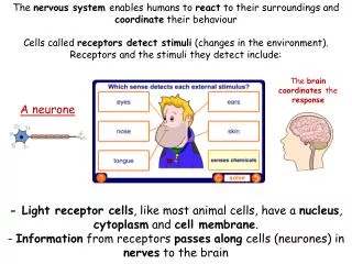
How do neurones transmit messages? 1) Sensory receptors (like rods and cones in the retina of the eye that detect changes in light intensity) convert the energy into a nerve impulse – a form of electrical energy. These nerve impulses are generated by changing the permeability of the neuron membrane. Neurones have specialised channel proteins that are specific to sodium and potassium ions. They are gated channels – which means they can be opened or closed to increase or decrease the permeability of the membrane. In addition, neurones have carrier proteins that actively transport Na+ ions out of the cell and K+ ions into the cell. Approximately 3 sodium ions are transported out for every 2 potassium brought in, so the inside of the cell is negatively charged relative to the outside. The membrane is said to be polarised – there is a potential difference across it. This is the resting potential and is usually about -60mV.
Sodium ion channels in the neurone membrane open. The membrane permeability to sodium increases and the sodium ions can diffuse across the membrane, down their concentration gradient into the cell. 2) Outside Cell The movement of ions across the membrane creates a change in the potential difference across the membrane. The inside of the cell becomes less negative compared to the outside – this is depolarisation. Na+ Na+ Na+ Na+ Na+ Inside Cell
There are channels in the membrane that respond to the depolarisations of the membrane. These are called voltage gated channels. They allow the passage of ions or charged particles. 3) A small depolarisation of the membrane will have no effect on these voltage gated channels. However, if the depolarisation is big enough, it will reach the threshold potential. This is a potential difference across the membrane of around -50mV. Na+ Outside Cell At this threshold potential, voltage-gated sodium ion channels will open. As more sodium ions flood into the neurone cell , its inside becomes more positively charged compared to its outside. Na+ Na+ When the potential difference across the membrane reaches +40mV, the inside of the cell is more positive than the outside. This is an action potential, and the neurone will transmit this along the axon, continuing to its end at the axon terminal. Na+ Na+ Na+ Na+ Inside Cell
4) At the point of the action potential, the sodium ion channels close, preventing them from entering. The potassium ion channels open, allowing the potassium ions to diffuse out of the cell. Outside Cell This makes the potential difference across the neurone cell membrane return to being negative – the inside of the cell is again more negative than the outside. This is called repolarisation K+ Na+ K+ The potential difference reaches more than -60mV, so the difference between the inside and outside of the cell is bigger than the resting potential. This is hyperpolarisation . The original potential difference will eventually be restored and the neurone returns to its resting state. K+ K+ K+ Inside Cell
After an action potential, the sodium and potassium ions are in the wrong places; there are potassium ions on the outside of the cell and there are sodium ions on the inside of the cell. These concentrations must be restored by the action of the sodium/potassium ion pumps. 5) There is a refractory period after each action potential. This is a time period in which the neurone’s cell membrane cannot be stimulated, allowing the cell to recover. There is a myelin sheath, an insulating layer of fatty material, around the axon. Sodium and potassium ions cannot diffuse through this. There are gaps between the Schwann cells which comprise the myelin sheath, and at these points, ions can move in and out. The gaps, called Nodes of Ranvier, mean that sodium ions diffuse along the neurone from one gap to the next. The action potential therefore seems to jump from one gap to the next – this is known as saltatory conduction.
Summary: An Action Potential Graph… 1. The membrane starts in its resting state – it is polarised, with the inside of the cell being -60mV compared to the outside. 2. The sodium ion channels open and sodium ions diffuse into the cell. The membrane depolarises, becoming less negative in relation to the outside. When the threshold potential reaches -50mV, voltage gated sodium ion channels will open, and more sodium ions enter. The inside of the cell is now positive in comparison to the outside, around +40mV. This is an action potential. 3. The sodium ion channels close and the potassium ion channels open. Potassium ions diffuse out of the cell, returning the potential difference across to being negative inside compared to the outside. This is repolarisation. 4. The potential difference goes slightly too low – too many K+ ions diffuse out. The potential difference will be restored, returning the membrane to its resting potential.
Transmission of Action Potentials in a Myelinated Neurone 3. This causes the ions to diffuse sideways, away from the region of increased concentration. This is a ‘local current’ – movement of charged Na+ ions in the cytoplasm of the neurone. 2. The concentration of sodium ions rises at the point where the sodium channels are open. 1. When an action potential occurs, the sodium ion channels open at a particular point along the neurone. This allows sodium ions to diffuse across the membrane. This is a positive feedback mechanism. 4. Further along the neurone are more voltage gated sodium ion channels. At rest, the potential difference across the membrane is -60mV. The movement of the sodium ions alters this, and as the potential difference across the membrane is reduced, the gates open. Sodium ions enter the neurone at a point further along the membrane. The action potential has moved along the neurone. In myelinated neurones, local currents are elongated and ions diffuse along the neurone form one node to the next. The action potentials ‘jump’ from node to node – this is saltatory conduction. The myelin sheath is an insulating layer. Sodium and potassium ions can’t diffuse through it, so action potentials can only occur at nodes of Ranvier where the axon membrane is exposed.
Unmyelinated neurones are found in the central nervous system (whereas myelinated neurones are found in the peripheral nervous system). The speed of conduction is much slower in an unmyelinated neurone – 0.5 m/s compared to 100m/s in a myelinated neurone. Multiple Sclerosis is a chronic autoimmune condition of the central nervous system. In a sufferer of multiple sclerosis, the body’s own immune system mistakes the myelin tissue for a foreign cell, and attacks it. This damages the myelin and strips it from the nerve fibres. This leaves scars called lesisons. This disrupts messages travelling along nerve fibres – they can slow down, become distorted, pass from one nerve fibre to another (short circuiting), or not get through at all. The loss of muscle coordination in people with MS is due to this degeneration of the myelin sheath in neurones involved in the movement of muscles.
All or Nothing… What does vary however is the frequency of the action potential and the number of neurones activated. A weak stimulus will create a low frequency of action potentials and few neurones will be stimulated. A strong stimulus will generate a high frequency of action potentials and lots of neurones will be stimulated. An action potential is an all or nothing response. A weak stimulus will not initiate an action potential: one will only be generated if the depolarisation of the receptor cell is large enough to reach the threshold potential of +40mV. The size of an action potential never changes: regardless of how strong the stimulus, the depolarisation always reaches +40mV. Synapses – “a gap between neurones” A synapse is a junction between two neurones. The neurones do not touch but are separated by a 20nm wide gap called a synaptic cleft. The presynaptic neurone releases a neurotransmitter substance that diffuses across the gap and generates a new action potential in the post synaptic neurone. The knob of the presynaptic neurone has many mitochondria to generate ATP for the active process involved; large amounts of smooth ER; lots of vesicles containing acetylcholine; voltage gated Ca2+ channels.
Transmission across a synapse… Acetylcholine Choline 1. Action potential causes calcium ion channels in the pre-synaptic neurone membrane to open. 2. Calcium ions diffuse into the pre-synaptic knob through the voltage gated channels. 3. Calcium causes vesicles containing a neurotransmitter, acetylcholine, to move to the pre-synaptic membrane. Acetate 6. Sodium diffuses into the neurone depolarising the membrane. If enough Na+ enters to reach the threshold level, this generates an action potential in the post synaptic neurone. 4. Vesicles fuse with the pre-synaptic membrane. Acetylcholine is released into the cleft by exocytosis. It diffuses across the synaptic cleft to the post synaptic membrane. 5. Acetylcholine binds to the receptor proteins on the post synaptic neurone, opening sodium channels. The enzyme acetylcholinesteraseis found in synaptic clefts. It breaks down acetylcholine into acetate and choline. Choline is taken back into the presynaptic neurone where it is combined with acetyl CoA to form acetylcholine once more.
The role of synapses… Impulses travel in only one direction at synapses. Vesicles with the neurotransmitter substance are present only in the pre synaptic neurone. Only the post synaptic neurones have receptors. Synapses allow for integration within the nervous system. One neurone can have several hundred synapses with other neurones. Lots of presynaptic neurones may converge to one post synaptic neurone, allowing signals from different parts of the nervous system to create the same response. One presynaptic neurone may diverge to several post synaptic neurones allowing one signal to be transmitted to several parts of the nervous system. Synapses increase the range of actions possible in response to a stimulus. If a synapse is excitatory, the neurotransmitter depolarises the membrane creating an action potential. If a synapse is inhibitory, the neurotransmitter hyperpolarises the membrane, so no action potential is formed. Summation is the interaction of many small potentials. Temporal Summation: One small action potential in the presynaptic neurone does not produce an action potential in the post synaptic neurone . A series of action potentials in the presynaptic neurone is required to generate an action potential in the post-synaptic neurone. This filters out low level stimuli. Spatial Summation is when several presynaptic neurones may each contribute to producing an action potential in the post synaptic neurone. Fatigue: continued stimulation results in the presynaptic neurone running out of transmitter, stopping an action potential. This filters out low level persistent stimuli. Memory and Learning: new synapses may form as a result of experience.